BD556547-BD细胞凋亡试剂盒FITC Annexin V Apoptosis Detection Kit I

【简单介绍】

ContentsAnnexin V-FITC, Propidium Iodide Staining Solution, Annexin V Binding BufferSize100 TestsRegulatory Status RUO
*,现货促销,*,咨询

【详细说明】

 Description

Apoptosis is a normal physiologic process which occurs during embryonic development as well as in maintenence of tissue homeostasis. The

apoptotic program is characterized by certain morphologic features, including loss of plasma membrane asymmetry and attachment,

condensation of the cytoplasm and nucleus, and internucleosomal cleavage of DNA. Loss of plasma membrane is one of the earliest features.

In apoptotic cells, the membrane phospholipid phosphatidylserine (PS) is translocated from the inner to the outer leaflet of the plasma

membrane, thereby exposing PS to the external cellular environment. Annexin V is a 35-36 kDa Ca2+ dependent phospholipid-binding

protein that has a high affinity for PS, and binds to cells with exposed PS. Annexin V may be conjugated to fluorochromes including FITC.

This format retains its high affinity for PS and thus serves as a sensitive probe for flow cytometric analysis of cells that are undergoing

apoptosis. Since externalization of PS occurs in the earlier stages of apoptosis, FITC Annexin V staining can identify apoptosis at an earlier

stage than assays based on nuclear changes such as DNA fragmentation.

FITC Annexin V staining precedes the loss of membrane integrity which accompanies the latest stages of cell death resulting from either

apoptotic or necrotic processes. Therefore, staining with FITC Annexin V is typically used in conjunction with a vital dye such as propidium

iodide (PI) or 7-Amino-Actinomycin (7-AAD) to allow the investigator to identify early apoptotic cells (PI negative, FITC Annexin V

positive). Viable cells with intact membranes exclude PI, wheras the membranes of dead and damaged cells are permeable to PI. For example,

cells that are considered viable are FITC Annexin V and PI negative; cells that are in early apoptosis are FITC Annexin V positive and PI

negative; and cells that are in late apoptosis or already dead are are both FITC Annexin V and PI positive. This assay does not distinguish

between cells that have undergone apoptotic death versus those that have died as a result of a necrotic pathway because in either case, the dead

cells will stain with both FITC Annexin V and PI. However, when apoptosis is measured over time, cells can be often tracked from FITC

Annexin V and PI negative (viable, or no measurable apoptosis), to FITC Annexin V positive and PI negative (early apoptosis, membrane

integrity is present) and finally to FITC Annexin V and PI positive (end stage apoptosis and death). The movement of cells through these three

stages suggests apoptosis. In contrast, a single observation indicating that cells are both FITC Annexin V and PI positive, in of itself, reveals

less information about the process by which the cells underwent their demise.

Preparation and Storage

Store undiluted at 4°C and protected from prolonged exposure to light. Do not freeze.

556547 Rev. 5 Page 1 of 3

Flow Cytometric Analysis of FITC Annexin V staining. Jurkat cells

(Human T-cell leukemia; ATCC TIB-152) were left untreated (top

panels) or treated for 4 hours with 12 μM campotothecin (bottom

panels). Cells were incubated with FITC Annexin V in a buffer

containing propidium iodide (PI) and analyzed by flow cytometry.

Untreated cells were primarily FITC Annexin V and PI negative,

indicating that they were viable and not undergoing apoptosis. After a

4 hour treatment (bottom panels), there were primarily two

populations of cells: Cells that were viable and not undergoing

apoptosis (FITC Annexin V and PI negative) and cells undergoing

apoptosis (FITC Annexin V positive and PI negative). A minor

population of cells were observed to be FITC Annexin V and PI

positive, indicating that they were in end stage apoptosis or already

dead.

Application Notes

Application

Flow cytometry Routinely Tested

Recommended Assay Procedure:

FITC Annexin V is a sensitive probe for identifying apoptotic cells, binding to negatively charged phospholipid surfaces (Kd of ~5 x 10^-2) with

a higher affinity for phosphatidylserine (PS) than most other phospholipids. FITC Annexin V binding is calcium dependent and defined calcium

and salt concentrations are required for optimal staining as described in the FITC Annexin V Staining Protocol. Investigators should note that

FITC Annexin V flow cytometric analysis on adherent cell types (e.g HeLa, NIH 3T3, etc.) is not routinely tested as specific membrane

damage may occur during cell detachment or harvesting. Methods for utilizing Annexin V for flow cytometry on adherent cell types,

however, have been previously reported (Casiola-Rosen et al. and van Engelend et al.).

INDUCTION OF APOPTOSIS BY CAMPTOTHECIN

The following protocol is provided as an illustration on how FITC Annexin V may be used on a cell line (Jurkat).

Materials

1. Prepare Camptothecin stock solution (Sigma-Aldrich Cat. No. C-9911): 1 mM in DMSO.

2. Jurkat T cells (ATCC TIB-152).

Procedure

1. Add Camptothecin (final conc. 4-6 μM) to 1 x 10^6 Jurkat cells.

2. Incubate the cells for 4-6 hr at 37°C.

3. Proceed with the FITC Annexin V Staining Protocol to measure apoptosis.

FITC ANNEXIN V STAINING PROTOCOL

FITC Annexin V is used to quantitatively determine the percentage of cells within a population that are actively undergoing apoptosis. It relies on

the property of cells to lose membrane asymmetry in the early phases of apoptosis. In apoptotic cells, the membrane phospholipid

phosphatidylserine (PS) is translocated from the inner leaflet of the plasma membrane to the outer leaflet, thereby exposing PS to the external

environment. Annexin V is a calcium-dependent phospholipid-binding protein that has a high affinity for PS, and is useful for identifying

apoptotic cells with exposed PS. Propidium Iodide (PI) is a standard flow cytometric viability probe and is used to distinguish viable from

nonviable cells. Viable cells with intact membranes exclude PI, whereas the membranes of dead and damaged cells are permeable to PI. Cells that

stain positive for FITC Annexin V and negative for PI are undergoing apoptosis. Cells that stain positive for both FITC Annexin V and PI are

either in the end stage of apoptosis, are undergoing necrosis, or are already dead. Cells that stain negative for both FITC Annexin V and PI are

alive and not undergoing measurable apoptosis.

556547 Rev. 5 Page 2 of 3

Reagents

1. FITC Annexin V (component no. 51-65874X): Use 5 μl per test.

2. Propidium Iodide (PI) (component no. 51-66211E) is a convenient, ready-to-use nucleic acid dye. Use 5 μl per test.

3. 10X Annexin V Binding Buffer (component no. 51-66121E): 0.1 M Hepes/NaOH (pH 7.4), 1.4 M NaCl, 25 mM CaCl2. For a 1X working

solution, dilute 1 part of the 10X Annexin V Binding Buffer to 9 parts of distilled water.

Staining

1. Wash cells twice with cold PBS and then resuspend cells in 1X Binding Buffer at a concentration of 1 x 10^6 cells/ml.

2. Transfer 100 μl of the solution (1 x 10^5 cells) to a 5 ml culture tube.

3. Add 5 μl of FITC Annexin V and 5 μl PI.

4. Gently vortex the cells and incubate for 15 min at RT (25°C) in the dark.

5. Add 400 μl of 1X Binding Buffer to each tube. Analyze by flow cytometry within 1 hr.

SUGGESTED CONTROLS FOR SETTING UP FLOW CYTOMETRY

The following controls are used to set up compensation and quadrants:

1. Unstained cells.

2. Cells stained with FITC Annexin V (no PI).

3. Cells stained with PI (no FITC Annexin V).

Other Staining Controls:

A cell line that can be easily induced to undergo apoptosis should be used to obtain positive control staining with FITC Annexin V and/or FITC

Annexin V and PI. It is important to note that the basal level of apoptosis and necrosis varies considerably within a population. Thus, even in the

absence of induced apoptosis, most cell populations will contain a minor percentage of cells that are positive for apoptosis (FITC Annexin V

positive, PI negative or FITC Annexin V positive, PI positive).

The untreated population is used to define the basal level of apoptotic and dead cells. The percentage of cells that have been induced to undergo

apoptosis is then determined by subtracting the percentage of apoptotic cells in the untreated population from percentage of apoptotic cells in the

treated population. Since cell death is the eventual outcome of cells undergoing apoptosis, cells in the late stages of apoptosis will have a damaged

membrane and stain positive for PI as well as for FITC Annexin V. Thus the assay does not distinguish between cells that have already undergone

an apoptotic cell death and those that have died as a result of necrotic pathway, because in either case the dead cells will stain with both FITC

Annexin V and PI.

Product Notices

1. Since applications vary, each investigator should titrate the reagent to obtain optimal results.

2. Source of all serum proteins is from USDA inspected abattoirs located in the United States.

Caution: Sodium azide yields highly toxic hydrazoic acid under acidic conditions. Dilute azide compounds in running water before

discarding to avoid accumulation of potentially explosive deposits in plumbing.

3.

4. Please refer to www.bdbiosciences.com/pharmingen/protocols for technical protocols.

References

Andree HA, Reuingsperger CP, Hauptmann R, Hemker HC, Hermens WT, Willems GM. Binding of vascular anticoagulant alpha (VAC alpha) to planar

phospholipid bilayers. J Biol Chem. 1990; 265(9):4923-4928. (Biology)

Casciola-Rosen L, Rosen A, Petri M, Schlissel M. Surface blebs on apoptotic cells are sites of enhanced procoagulant activity: implications for coagulation events

and antigenic spread in systemic lupus erythematosus. Proc Natl Acad Sci U S A. 1996; 93(4):1624-1629. (Biology)

Homburg CH, de Haas M, von dem Borne AE, Verhoeven AJ, Reuingsperger CP, Roos D. Human neutrophils lose their surface Fc gamma RIII and acquire

Annexin V binding sites during apoptosis in vitro. Blood. 1995; 85(2):532-540. (Biology)

Koopman G, Reuingsperger CP, Kuijten GA, Keehnen RM, Pals ST, van Oers MH. Annexin V for flow cytometric detection of phosphatidylserine expression on

B cells undergoing apoptosis. Blood. 1994; 84(5):1415-1420. (Biology)

Martin SJ, Reuingsperger CP, McGahon AJ, et al. Early redistribution of plasma membrane phosphatidylserine is a general feature of apoptosis regardless of

the initiating stimulus: inhibition by overexpression of Bcl-2 and Abl. J Exp Med. 1995; 182(5):1545-1556. (Biology)

O'Brien MC, Bolton WE. Comparison of cell viability probes compatible with fixation and permeabilization for combined surface and intracellular staining in flow

cytometry. Cytometry. 1995; 19(3):243-255. (Biology)

Raynal P, Pollard HB. Annexins: the problem of assessing the biological role for a gene family of multifunctional calcium- and phospholipid-binding proteins.

Biochim Biophys Acta. 1994; 1197(1):63-93. (Biology)

Schmid I, Krall WJ, Uittenbogaart CH, Braun J, Giorgi JV. Dead cell discrimination with 7-amino-actinomycin D in combination with dual color immunofluorescence

in single laser flow cytometry. Cytometry. 1992; 13(2):204-208. (Biology)

van Engeland M, Ramaekers FC, Schutte B, Reuingsperger CP. A novel assay to measure loss of plasma membrane asymmetry during apoptosis of adherent

cells in culture. Cytometry. 1996; 24(2):131-139. (Biology)

Vermes I, Haanen C, Steffens-Nakken H, Reuingsperger C. A novel assay for apoptosis. Flow cytometric detection of phosphatidylserine expression on early

apoptotic cells using fluorescein labelled Annexin V. J Immunol Methods. 1995; 184(1):39-51. (Biology)

556547


Cell Meter 磷脂酰丝氨酸凋亡检测试剂盒 蓝色荧光,适合微孔板检测-AAT Bioquest荧光染料

上海金畔生物科技有限公司代理AAT Bioquest荧光染料全线产品,欢迎访问AAT Bioquest荧光染料官网了解更多信息。
Cell Meter 磷脂酰丝氨酸凋亡检测试剂盒 蓝色荧光,适合微孔板检测 价格 2823
产品规格

100 Tests

产品货号

Cell Meter 磷脂酰丝氨酸凋亡检测试剂盒 蓝色荧光,适合微孔板检测

产品参数
Ex (nm) Em (nm)
分子量 溶剂
存储条件
产品概述

我们的Cell Meter™检测试剂盒是一套用于监测细胞活性的试剂盒。有多种参数可用于监测细胞活力。该特定试剂盒旨在通过测量磷脂酰丝氨酸(PS)的转运来监测细胞凋亡。在细胞凋亡中,PS转移到质膜的外部小叶。磷脂酰丝氨酸在细胞表面的出现是细胞凋亡的初始/中间阶段的通用指标,可以在观察形态变化之前进行检测。该特定试剂盒旨在通过测量磷脂酰丝氨酸(PS)的转运来监测细胞凋亡。该试剂盒使用我们专有的基于荧光小分子的Apopxin™PS探针,该探针以比膜联蛋白V(Kd <10 nM)高得多的亲和力特异性结合PS。与膜PS结合后具有蓝色荧光。它可以以酶标仪,显微镜和流式细胞仪的形式使用,而大多数其他商业凋亡检测试剂盒只能与显微镜或流式细胞仪平台一起使用。


适用仪器


荧光酶标仪  
Ex: 405 nm
Em: 450 nm
Cutoff: 420 nm
推荐孔板: 黑色透明底板
读取模式: 底读模式
实验方案

样品实验方案

简要概述

1.用测试化合物制备细胞(100 µL /孔/ 96孔板或25 µL /孔/ 384孔板)
2.加入等量的Apopxin™Violet 450工作溶液
3.在室温下孵育1小时
4.观察Ex / Em = 405/450 nm(截止= 420 nm)的荧光强度(底部读取模式)或使用装有紫罗兰色滤光片的荧光显微镜


溶液配制

工作溶液配制

将10μL100X Apopxin™紫罗兰色450(组分A)添加到1 mL的测定缓冲液(组分B)中,并充分混合以制成Apopxin™紫罗兰色450工作溶液。 


操作步骤

1.通过向PBS或所需的缓冲液中加入10X /孔(96孔板)或2.5 µL /孔(384孔板)的10X测试化合物储备溶液来处理细胞。对于空白孔(不含细胞的培养基),添加相同量的化合物缓冲液。

2.将细胞板在5%CO2、37°C​​的培养箱中孵育所需的时间(对于用星形孢菌素处理的Jurkat细胞需要4-6小时)以诱导凋亡。注意:由于在405 nm激发时,从380至490 nm的宽发射光谱,某些化合物(例如喜树碱)可能会产生假阳性反应。

3.在每个孔中添加100 µL /孔(96孔板)或25 µL /孔(384孔板)的Apopxin™Violet 450工作溶液。

4.在避光条件下,于室温下孵育细胞板至少1小时。

5.以800 rpm的速度离心细胞板(特别是非粘附细胞)2分钟(制动)。

6.使用荧光酶标仪(底部读取模式)在Ex / Em = 405/450 nm(截止= 420 nm)或使用带有紫色滤光片的荧光显微镜对图像池进行荧光强度监测。

Cell Meter 磷脂酰丝氨酸凋亡检测试剂盒 蓝色荧光,适合微孔板检测

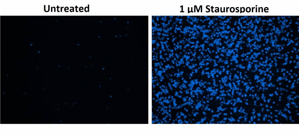
图1.用Apopxin™Violet 450偶联物染色的HeLa细胞的荧光图像。 Jurkat细胞在37℃下不使用(左)或用1μM星形孢菌素(右)处理4小时。 使用带有紫色滤光片组的显微镜(激发= 405nm)测量荧光强度。

参考文献

Physiological effects of the herbicide glyphosate on the cyanobacterium Microcystis aeruginosa
Authors: Wu, Liang and Qiu, Zhihao and Zhou, Ya and Du, Yuping and Liu, Chaonan and Ye, Jing and Hu, Xiaojun
Journal: Aquatic Toxicology (2016): 72–79

Tumor-selective mitochondrial network collapse induced by atmospheric gas plasma-activated medium.
Authors: Saito, Kosuke and Asai, Tomohiko and Fujiwara, Kyoko and Sahara, Junki and Koguchi, Haruhisa and Fukuda, Noboru and Suzuki-Karasaki, Miki and Soma, Masayoshi and Suzuki-Karasaki, Yoshihiro
Journal: Oncotarget (2016)

Inhibition of malignant phenotypes of human osteosarcoma cells by a gene silencer, a pyrrole–imidazole polyamide, which targets an E-box motif
Authors: Taniguchi, Masashi and Fujiwara, Kyoko and Nakai, Yuji and Ozaki, Toshinori and Koshikawa, Nobuko and Toshio, Kojima and Kataba, Motoaki and Oguni, Asako and Matsuda, Hiroyuki and Yoshida, Yukihiro and others
Journal: FEBS open bio (2014): 328–334

Detection of apoptosis based on the interaction between annexin V and phosphatidylserine
Authors: Liu T, Zhu W, Yang X, Chen L, Yang R, Hua Z, Li G.
Journal: Anal Chem (2009): 2410

Evaluation of cell surface expression of phosphatidylserine in ovarian carcinoma effusions using the annexin-V/7-AAD assay: clinical relevance and comparison with other apoptosis parameters
Authors: Dong HP, Holth A, Kleinberg L, Ruud MG, Elstr and MB, Trope CG, Davidson B, Risberg B.
Journal: Am J Clin Pathol (2009): 756

Mobilization of lysosomal calcium regulates the externalization of phosphatidylserine during apoptosis
Authors: Mirnikjoo B, Balasubramanian K, Schroit AJ.
Journal: J Biol Chem (2009): 6918

Peptidic targeting of phosphatidylserine for the MRI detection of apoptosis in atherosclerotic plaques
Authors: Burtea C, Laurent S, Lancelot E, Ballet S, Murariu O, Rousseaux O, Port M, V and er Elst L, Corot C, Muller RN.
Journal: Mol Pharm (2009): 1903

Suicidal membrane repair regulates phosphatidylserine externalization during apoptosis
Authors: Mirnikjoo B, Balasubramanian K, Schroit AJ.
Journal: J Biol Chem (2009): 22512

Trivalent methylated arsenical-induced phosphatidylserine exposure and apoptosis in platelets may lead to increased thrombus formation
Authors: Bae ON, Lim KM, Noh JY, Chung SM, Kim SH, Chung JH.
Journal: Toxicol Appl Pharmacol (2009): 144

Discovery of a phosphatidylserine-recognizing peptide and its utility in molecular imaging of tumour apoptosis
Authors: Thapa N, Kim S, So IS, Lee BH, Kwon IC, Choi K, Kim IS.
Journal: J Cell Mol Med (2008): 1649

Cell Meter 磷脂酰丝氨酸凋亡检测试剂盒 绿色荧光,适合微孔板检测-AAT Bioquest荧光染料

上海金畔生物科技有限公司代理AAT Bioquest荧光染料全线产品,欢迎访问AAT Bioquest荧光染料官网了解更多信息。
Cell Meter 磷脂酰丝氨酸凋亡检测试剂盒 绿色荧光,适合微孔板检测 价格 2823
产品规格

100 Tests

产品货号

Cell Meter 磷脂酰丝氨酸凋亡检测试剂盒 绿色荧光,适合微孔板检测

产品参数
Ex (nm) Em (nm)
分子量 溶剂
存储条件
产品概述

Cell Meter检测试剂盒是一类用于检测细胞功能的系列工具,包括细胞活性、细胞毒性、细胞凋亡、细胞膜电位以及细胞周期等方面的指标。每种检测方案均能提供不同荧光颜色的检测方案。这些检测方案为从多角度研究细胞功能活动提供了一种十分有效的方法。Cell Meter磷脂酰丝氨酸凋亡检测试剂盒 *绿色荧光,适合微孔板检测*是通过检测PS的翻转来测定细胞凋亡。在凋亡细胞中,PS从胞内移位到胞外表面,因此PS出现在细胞的外表面可以作为细胞凋亡的起始或者中间阶段的通用指示器,也可以通过其形态学改变来测定。该试剂盒使用我们独有的绿色荧光Apopxin PS传感器特异性的结合PS,其亲和性高于Annexin V(Kd<10nM)。该试剂盒使用的PS传感器结合膜上的PS后,发出绿色的荧光。因为与PS的高亲和性,该试剂盒比其它基于Annexin-V,仅能用荧光显微镜和流式检测凋亡的试剂盒更稳定,并且本试剂盒除上述两种仪器外还能使用荧光酶标仪。

点击查看细胞样品制备


适用仪器


荧光酶标仪  
Ex: 490 nm
Em: 525 nm
Cutoff: 515 nm
推荐孔板: 黑色透明底板
读取模式: 底读模式
实验方案

样品实验方案

简要概述

1.用测试化合物制备细胞(100 µL /孔/ 96孔板或25 µL /孔/ 384孔板)
2.加入等量的Apopxin™Green工作溶液
3.在室温下孵育1小时
4.在Ex / Em = 490/525 nm(截止= 515 nm)或带有FITC滤光片的荧光显微镜下监控荧光强度(底部读取模式)


溶液配制

工作溶液配制

将10 µL 100X Apopxin™Green(组分A)添加到1 mL的测定缓冲液(组分B)中,并充分混合以制成Apopxin™Green工作溶液。


操作步骤

1.通过向细胞制备缓冲液中加入10XL /孔(96孔板)的10X测试化合物溶液,以所需的测试化合物处理细胞,总体积为100μL/孔。对于384孔板,请在细胞制备缓冲液中使用5 µL /孔的5X测试化合物溶液,总体积为25 µL /孔。对于空白孔(不含细胞的培养基),添加相同量的化合物缓冲液。

2.将细胞板在5%CO2、37°C​​的培养箱中孵育所需的时间(对于喜树碱处理过的Jurkat细胞,需要4-6小时)以诱导凋亡。

3.在每个孔中添加100 µL /孔(96孔板)或25 µL /孔(384孔板)的Apopxin™Green工作溶液。

4.在避光条件下,于室温下孵育细胞板至少1小时。

5.以800 rpm的速度离心细胞板(特别是非粘附细胞)2分钟(制动)。

6.使用荧光酶标仪(Ext / Em = 490/525 nm(Cutoff = 515 nm))或使用带有FITC滤光片的荧光显微镜的图像池监控荧光强度。


图示

Cell Meter 磷脂酰丝氨酸凋亡检测试剂盒 绿色荧光,适合微孔板检测

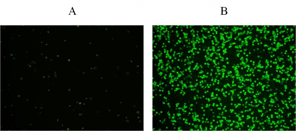
图1.在Costar黑色壁/透明底部96孔板中,用Cell Meter™磷脂酰丝氨酸凋亡检测试剂盒染色的Jurkat细胞图像。 A:未经处理的对照细胞。 B:用20 µM喜树碱处理5小时的细胞。

参考文献

Physiological effects of the herbicide glyphosate on the cyanobacterium Microcystis aeruginosa
Authors: Wu, Liang and Qiu, Zhihao and Zhou, Ya and Du, Yuping and Liu, Chaonan and Ye, Jing and Hu, Xiaojun
Journal: Aquatic Toxicology (2016): 72–79

Tumor-selective mitochondrial network collapse induced by atmospheric gas plasma-activated medium.
Authors: Saito, Kosuke and Asai, Tomohiko and Fujiwara, Kyoko and Sahara, Junki and Koguchi, Haruhisa and Fukuda, Noboru and Suzuki-Karasaki, Miki and Soma, Masayoshi and Suzuki-Karasaki, Yoshihiro
Journal: Oncotarget (2016)

Inhibition of malignant phenotypes of human osteosarcoma cells by a gene silencer, a pyrrole–imidazole polyamide, which targets an E-box motif
Authors: Taniguchi, Masashi and Fujiwara, Kyoko and Nakai, Yuji and Ozaki, Toshinori and Koshikawa, Nobuko and Toshio, Kojima and Kataba, Motoaki and Oguni, Asako and Matsuda, Hiroyuki and Yoshida, Yukihiro and others
Journal: FEBS open bio (2014): 328–334

Detection of apoptosis based on the interaction between annexin V and phosphatidylserine
Authors: Liu T, Zhu W, Yang X, Chen L, Yang R, Hua Z, Li G.
Journal: Anal Chem (2009): 2410

Evaluation of cell surface expression of phosphatidylserine in ovarian carcinoma effusions using the annexin-V/7-AAD assay: clinical relevance and comparison with other apoptosis parameters
Authors: Dong HP, Holth A, Kleinberg L, Ruud MG, Elstr and MB, Trope CG, Davidson B, Risberg B.
Journal: Am J Clin Pathol (2009): 756

Mobilization of lysosomal calcium regulates the externalization of phosphatidylserine during apoptosis
Authors: Mirnikjoo B, Balasubramanian K, Schroit AJ.
Journal: J Biol Chem (2009): 6918

Peptidic targeting of phosphatidylserine for the MRI detection of apoptosis in atherosclerotic plaques
Authors: Burtea C, Laurent S, Lancelot E, Ballet S, Murariu O, Rousseaux O, Port M, V and er Elst L, Corot C, Muller RN.
Journal: Mol Pharm (2009): 1903

Suicidal membrane repair regulates phosphatidylserine externalization during apoptosis
Authors: Mirnikjoo B, Balasubramanian K, Schroit AJ.
Journal: J Biol Chem (2009): 22512

Trivalent methylated arsenical-induced phosphatidylserine exposure and apoptosis in platelets may lead to increased thrombus formation
Authors: Bae ON, Lim KM, Noh JY, Chung SM, Kim SH, Chung JH.
Journal: Toxicol Appl Pharmacol (2009): 144

Discovery of a phosphatidylserine-recognizing peptide and its utility in molecular imaging of tumour apoptosis
Authors: Thapa N, Kim S, So IS, Lee BH, Kwon IC, Choi K, Kim IS.
Journal: J Cell Mol Med (2008): 1649

Cell Meter 磷脂酰丝氨酸凋亡检测试剂盒 红色荧光,适合微孔板检测-AAT Bioquest荧光染料

上海金畔生物科技有限公司代理AAT Bioquest荧光染料全线产品,欢迎访问AAT Bioquest荧光染料官网了解更多信息。
Cell Meter 磷脂酰丝氨酸凋亡检测试剂盒 红色荧光,适合微孔板检测 价格 2823
产品规格

100 Tests

产品货号

Cell Meter 磷脂酰丝氨酸凋亡检测试剂盒 红色荧光,适合微孔板检测

产品参数
Ex (nm) Em (nm)
分子量 溶剂
存储条件
产品概述

Cell Meter检测试剂盒是一类用于检测细胞功能的系列工具,包括细胞活性、细胞毒性、细胞凋亡、细胞膜电位以及细胞周期等方面的指标。每种检测方案均能提供不同荧光颜色的检测方案。这些高效的检测方案为从多角度研究细胞功能活动提供了一种十分有效的方法。Cell Meter磷脂酰丝氨酸凋亡检测试剂盒 *红色荧光,适合微孔板检测*是通过检测PS的翻转来测定细胞凋亡。在凋亡细胞中,PS从胞内移位到胞外表面,因此PS出现在细胞的外表面可以作为细胞凋亡的起始或者中间阶段的通用指示器,也可以通过其形态学改变来测定。该试剂盒使用我们独有的红色荧光Apopxin PS传感器特异性的结合PS,其亲和性高于Annexin V(Kd<10nM)。该试剂盒使用的PS传感器结合膜上的PS后,发出红色的荧光。因为与PS的高亲和性,该试剂盒比其它基于Annexin-V,仅能用荧光显微镜和流式检测凋亡的试剂盒更稳定,并且本试剂盒除上述两种仪器外还能使用荧光酶标仪。

点击查看细胞样品配制


适用仪器


荧光酶标仪  
Ex: 590 nm
Em: 630 nm
Cutoff: 610 nm
推荐孔板: 黑色透明底板
读取模式: 底读模式
实验方案

样品实验方案

简要概述

1.用测试化合物制备细胞(100 µL /孔/ 96孔板或25 µL /孔/ 384孔板)
2.加入等量的Apopxin™Red工作溶液
3.在室温下孵育1小时
4.监视Ex / Em = 590/630 nm(截止= 610 nm)的荧光强度(底部读取模式)或使用带有Texas Red滤光片的荧光显微镜


溶液配制

工作溶液配制

将10 µL Apopxin™Red(组分A)加入1 mL的测定缓冲液(组分B)中,并充分混合以制成Apopxin™Red工作溶液。


操作步骤

1.通过向PBS或所需的缓冲液中加入10X /孔(96孔板)或2.5 µL /孔(384孔板)的10X测试化合物储备溶液来处理细胞。 对于空白孔(不含细胞的培养基),添加相同量的化合物缓冲液。

2.将细胞板在5%CO2、37°C的培养箱中孵育所需的时间(对于喜树碱处理过的Jurkat细胞,需要4-6小时)以诱导凋亡。

3.在每个孔中加入100 µL /孔(96孔板)或25 µL /孔(384孔板)的Apopxin™Red工作溶液。

4.在避光条件下,于室温下孵育细胞板至少1小时。

5.以800 rpm的速度离心细胞板(特别是非粘附细胞)2分钟(制动)。

6.使用荧光酶标仪(Ext / Em = 590/630 nm(截止= 610 nm))或使用带有TexasRed®滤光片的荧光显微镜对图像池进行荧光强度监测。


图示

Cell Meter 磷脂酰丝氨酸凋亡检测试剂盒 红色荧光,适合微孔板检测

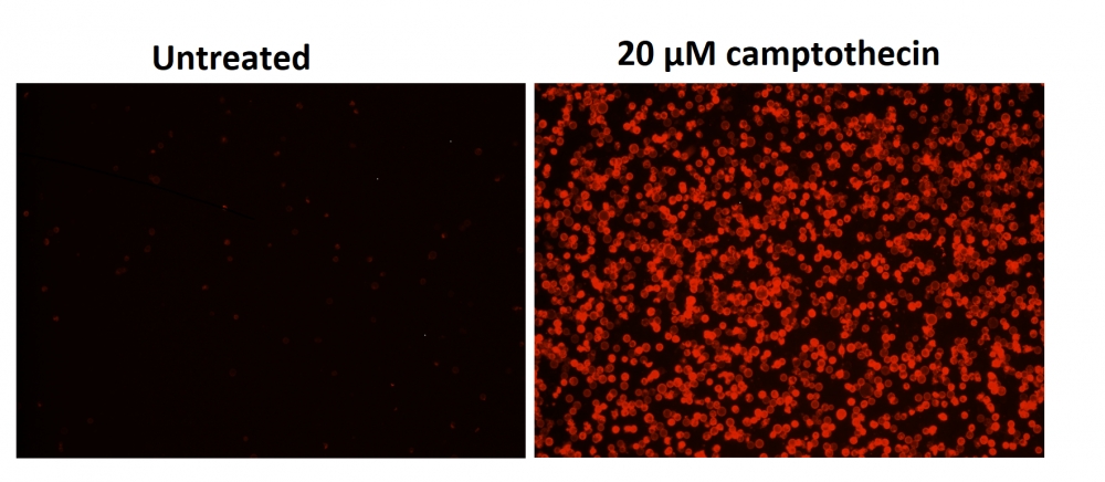
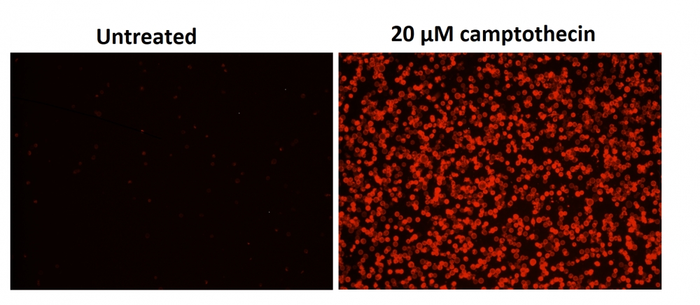
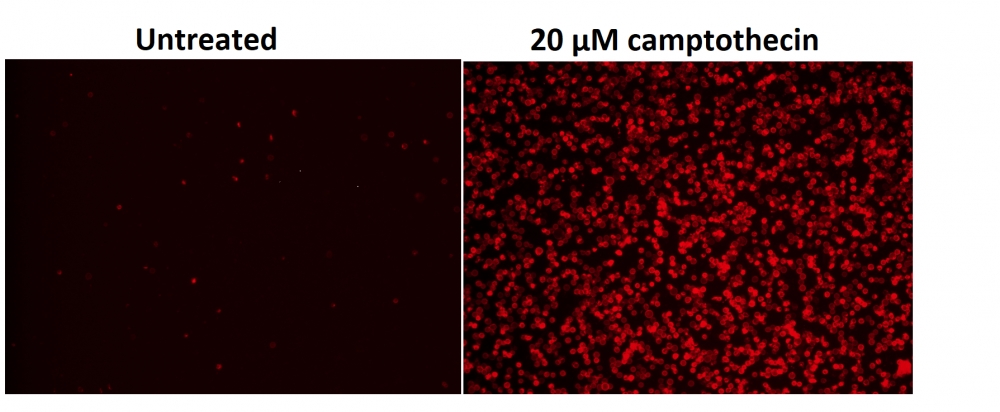
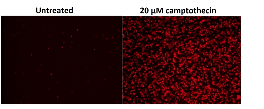
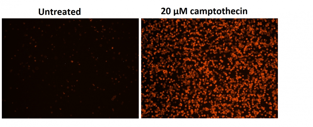
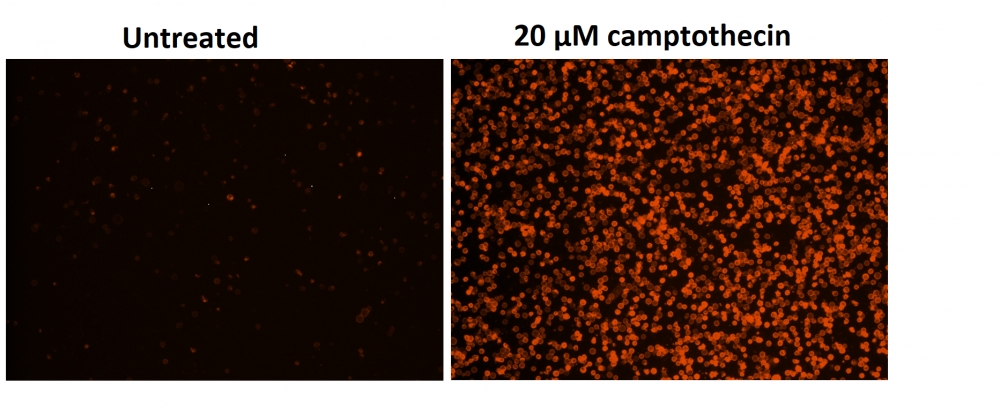
图1.用Cell Meter™磷脂酰丝氨酸凋亡检测试剂盒染色的Costar黑壁/透明底部96孔板中Jurkat细胞的荧光图像。 不使用(左)或用20μM喜树碱(右)处理Jurkat细胞5小时。 使用具有Texas Red通道的荧光显微镜测量荧光强度。


参考文献

Physiological effects of the herbicide glyphosate on the cyanobacterium Microcystis aeruginosa
Authors: Wu, Liang and Qiu, Zhihao and Zhou, Ya and Du, Yuping and Liu, Chaonan and Ye, Jing and Hu, Xiaojun
Journal: Aquatic Toxicology (2016): 72–79

Tumor-selective mitochondrial network collapse induced by atmospheric gas plasma-activated medium.
Authors: Saito, Kosuke and Asai, Tomohiko and Fujiwara, Kyoko and Sahara, Junki and Koguchi, Haruhisa and Fukuda, Noboru and Suzuki-Karasaki, Miki and Soma, Masayoshi and Suzuki-Karasaki, Yoshihiro
Journal: Oncotarget (2016)

Inhibition of malignant phenotypes of human osteosarcoma cells by a gene silencer, a pyrrole–imidazole polyamide, which targets an E-box motif
Authors: Taniguchi, Masashi and Fujiwara, Kyoko and Nakai, Yuji and Ozaki, Toshinori and Koshikawa, Nobuko and Toshio, Kojima and Kataba, Motoaki and Oguni, Asako and Matsuda, Hiroyuki and Yoshida, Yukihiro and others
Journal: FEBS open bio (2014): 328–334

Detection of apoptosis based on the interaction between annexin V and phosphatidylserine
Authors: Liu T, Zhu W, Yang X, Chen L, Yang R, Hua Z, Li G.
Journal: Anal Chem (2009): 2410

Evaluation of cell surface expression of phosphatidylserine in ovarian carcinoma effusions using the annexin-V/7-AAD assay: clinical relevance and comparison with other apoptosis parameters
Authors: Dong HP, Holth A, Kleinberg L, Ruud MG, Elstr and MB, Trope CG, Davidson B, Risberg B.
Journal: Am J Clin Pathol (2009): 756

Mobilization of lysosomal calcium regulates the externalization of phosphatidylserine during apoptosis
Authors: Mirnikjoo B, Balasubramanian K, Schroit AJ.
Journal: J Biol Chem (2009): 6918

Peptidic targeting of phosphatidylserine for the MRI detection of apoptosis in atherosclerotic plaques
Authors: Burtea C, Laurent S, Lancelot E, Ballet S, Murariu O, Rousseaux O, Port M, V and er Elst L, Corot C, Muller RN.
Journal: Mol Pharm (2009): 1903

Suicidal membrane repair regulates phosphatidylserine externalization during apoptosis
Authors: Mirnikjoo B, Balasubramanian K, Schroit AJ.
Journal: J Biol Chem (2009): 22512

Trivalent methylated arsenical-induced phosphatidylserine exposure and apoptosis in platelets may lead to increased thrombus formation
Authors: Bae ON, Lim KM, Noh JY, Chung SM, Kim SH, Chung JH.
Journal: Toxicol Appl Pharmacol (2009): 144

Discovery of a phosphatidylserine-recognizing peptide and its utility in molecular imaging of tumour apoptosis
Authors: Thapa N, Kim S, So IS, Lee BH, Kwon IC, Choi K, Kim IS.
Journal: J Cell Mol Med (2008): 1649

Cell Meter 磷脂酰丝氨酸凋亡检测试剂盒 深红色荧光,适合微孔板检测-AAT Bioquest荧光染料

上海金畔生物科技有限公司代理AAT Bioquest荧光染料全线产品,欢迎访问AAT Bioquest荧光染料官网了解更多信息。
Cell Meter 磷脂酰丝氨酸凋亡检测试剂盒 深红色荧光,适合微孔板检测 价格 2823
产品规格

100 Tests

产品货号

Cell Meter 磷脂酰丝氨酸凋亡检测试剂盒 深红色荧光,适合微孔板检测

产品参数
Ex (nm) Em (nm)
分子量 溶剂
存储条件
产品概述

Cell Meter检测试剂盒是一类用于检测细胞功能的系列工具,包括细胞活性、细胞毒性、细胞凋亡、细胞膜电位以及细胞周期等方面的指标。每种检测方案均能提供不同荧光颜色的检测方案。这些高效的检测方案为从多角度研究细胞功能活动提供了一种十分有效的方法。Cell Meter磷脂酰丝氨酸凋亡检测试剂盒 *深红色荧光,适合微孔板检测*是通过检测PS的翻转来测定细胞凋亡。在凋亡细胞中,PS从胞内移位到胞外表面,因此PS出现在细胞的外表面可以作为细胞凋亡的起始或者中间阶段的通用指示器,也可以通过其形态学改变来测定。该试剂盒使用我们独有的深红色荧光Apopxin PS传感器特异性的结合PS,其亲和性高于Annexin V(Kd<10nM)。该试剂盒使用的PS传感器结合膜上的PS后,发出深红色的荧光。因为与PS的高亲和性,该试剂盒比其它基于Annexin-V,仅能用荧光显微镜和流式检测凋亡的试剂盒更稳定,并且本试剂盒除上述两种仪器外还能使用荧光酶标仪。

点击查看细胞样品配制


适用仪器


荧光酶标仪  
Ex: 650 nm
Em: 680 nm
Cutoff: 665 nm
推荐孔板: 黑色透明底板
读取模式: 底读模式
实验方案

样品实验方案

简要概述

1.用测试化合物制备细胞(100 µL /孔/ 96孔板或25 µL /孔/ 384孔板)
2.加入等量的Apopxin™深红色工作溶液
3.在室温下孵育1小时
4.在Ex / Em = 650/680 nm(截止= 665 nm)或使用Cy5滤光片的荧光显微镜下监控荧光强度(底部读取模式)


溶液配制

工作溶液配制

将10μLApopxin™深红色(组分A)添加到1 mL的测定缓冲液(组分B)中,并充分混合以制成Apopxin™深红色工作溶液。


操作步骤

1.通过向PBS或所需的缓冲液中加入10X /孔(96孔板)或2.5 µL /孔(384孔板)的10X测试化合物储备溶液来处理细胞。 对于空白孔(不含细胞的培养基),添加相同量的化合物缓冲液。

2.将细胞板在5%CO2、37°C的培养箱中孵育所需的时间(对于喜树碱处理过的Jurkat细胞,需要4-6小时)以诱导凋亡。

3.在每个孔中加入100 µL /孔(96孔板)或25 µL /孔(384孔板)的Apopxin™深红色工作溶液。

4.在避光条件下,于室温下孵育细胞板至少1小时。

5.以800 rpm的速度离心细胞板(特别是非贴壁细胞)2分钟(制动)。

6.使用荧光酶标仪(Ext / Em = 650/680 nm(截止= 665 nm))或使用带有Cy5®滤光片的荧光显微镜对图像池进行荧光强度监测。


图示

Cell Meter 磷脂酰丝氨酸凋亡检测试剂盒 深红色荧光,适合微孔板检测

图1.用Cell Meter™磷脂酰丝氨酸凋亡检测试剂盒染色的Costar黑壁/透明底部96孔板中Jurkat细胞的荧光图像。 不使用(左)或用20μM喜树碱(右)处理Jurkat细胞5小时。 使用具有通道的荧光显微镜测量荧光强度。

参考文献

Physiological effects of the herbicide glyphosate on the cyanobacterium Microcystis aeruginosa
Authors: Wu, Liang and Qiu, Zhihao and Zhou, Ya and Du, Yuping and Liu, Chaonan and Ye, Jing and Hu, Xiaojun
Journal: Aquatic Toxicology (2016): 72–79

Tumor-selective mitochondrial network collapse induced by atmospheric gas plasma-activated medium.
Authors: Saito, Kosuke and Asai, Tomohiko and Fujiwara, Kyoko and Sahara, Junki and Koguchi, Haruhisa and Fukuda, Noboru and Suzuki-Karasaki, Miki and Soma, Masayoshi and Suzuki-Karasaki, Yoshihiro
Journal: Oncotarget (2016)

Inhibition of malignant phenotypes of human osteosarcoma cells by a gene silencer, a pyrrole–imidazole polyamide, which targets an E-box motif
Authors: Taniguchi, Masashi and Fujiwara, Kyoko and Nakai, Yuji and Ozaki, Toshinori and Koshikawa, Nobuko and Toshio, Kojima and Kataba, Motoaki and Oguni, Asako and Matsuda, Hiroyuki and Yoshida, Yukihiro and others
Journal: FEBS open bio (2014): 328–334

Detection of apoptosis based on the interaction between annexin V and phosphatidylserine
Authors: Liu T, Zhu W, Yang X, Chen L, Yang R, Hua Z, Li G.
Journal: Anal Chem (2009): 2410

Evaluation of cell surface expression of phosphatidylserine in ovarian carcinoma effusions using the annexin-V/7-AAD assay: clinical relevance and comparison with other apoptosis parameters
Authors: Dong HP, Holth A, Kleinberg L, Ruud MG, Elstr and MB, Trope CG, Davidson B, Risberg B.
Journal: Am J Clin Pathol (2009): 756

Mobilization of lysosomal calcium regulates the externalization of phosphatidylserine during apoptosis
Authors: Mirnikjoo B, Balasubramanian K, Schroit AJ.
Journal: J Biol Chem (2009): 6918

Peptidic targeting of phosphatidylserine for the MRI detection of apoptosis in atherosclerotic plaques
Authors: Burtea C, Laurent S, Lancelot E, Ballet S, Murariu O, Rousseaux O, Port M, V and er Elst L, Corot C, Muller RN.
Journal: Mol Pharm (2009): 1903

Suicidal membrane repair regulates phosphatidylserine externalization during apoptosis
Authors: Mirnikjoo B, Balasubramanian K, Schroit AJ.
Journal: J Biol Chem (2009): 22512

Trivalent methylated arsenical-induced phosphatidylserine exposure and apoptosis in platelets may lead to increased thrombus formation
Authors: Bae ON, Lim KM, Noh JY, Chung SM, Kim SH, Chung JH.
Journal: Toxicol Appl Pharmacol (2009): 144

Discovery of a phosphatidylserine-recognizing peptide and its utility in molecular imaging of tumour apoptosis
Authors: Thapa N, Kim S, So IS, Lee BH, Kwon IC, Choi K, Kim IS.
Journal: J Cell Mol Med (2008): 1649

Cell Meter 磷脂酰丝氨酸凋亡检测试剂盒 橙色荧光,适合酶标仪检测-AAT Bioquest荧光染料

上海金畔生物科技有限公司代理AAT Bioquest荧光染料全线产品,欢迎访问AAT Bioquest荧光染料官网了解更多信息。
Cell Meter 磷脂酰丝氨酸凋亡检测试剂盒 橙色荧光,适合酶标仪检测 价格 2823
产品规格

100 Tests

产品货号

Cell Meter 磷脂酰丝氨酸凋亡检测试剂盒 橙色荧光,适合酶标仪检测

产品参数
Ex (nm) Em (nm)
分子量 溶剂
存储条件
产品概述

Cell Meter检测试剂盒是一类用于检测细胞功能的系列工具,该特定试剂盒旨在通过测量磷脂酰丝氨酸(PS)的转运来检测细胞凋亡。在细胞凋亡中,PS转移到质膜的外部小叶。磷脂酰丝氨酸在细胞表面的出现是细胞凋亡的初始/中间阶段的通用指标,可以在观察形态变化之前进行检测。该试剂盒使用我们专有的橙色荧光Apopxin PS传感器,该传感器特异性结合PS,其亲和力远高于Annexin V(Kd <10 nM)。该试剂盒中使用的PS传感器与膜PS结合后具有橙色荧光。它的光谱性质几乎与Cy3®或AlexaFluor®555的光谱性质相同,使其易于用配备有Cy3®或AlexaFluor®555光源和滤光片的普通荧光仪器(Cy3®或AlexaFluor®555分别是GE Healthcare和Invitrogen的商标)进行实验。由于它对PS的亲和力大大增强,因此该试剂盒比仅与显微镜或流式细胞仪平台一起使用的其他基于膜联蛋白V的商业化凋亡试剂盒为强大。除显微镜和流式细胞仪平台外,该试剂盒还可与荧光酶标仪一起使用。

点击查看细胞样品配制


适用仪器


荧光酶标仪  
Ex: 540 nm
Em: 590 nm
Cutoff: 570 nm
推荐孔板: 黑色透明底板
读取模式: 底读模式
实验方案

样品实验方案

简要概述

  1. 用测试化合物制备细胞(100 µL /孔/ 96孔板或25 µL /孔/ 384孔板)
  2. 加入等量的Apopxin Orange工作溶液
  3. 在室温下孵育1小时
  4. 在Ex / Em = 540/590 nm(截止= 570 nm)或使用Cy3滤光片的荧光显微镜下检测荧光强度(底部读取模式)


溶液配制

工作溶液配制

将10μL的100X Apopxin 橙色(组分A)添加到1 mL的测定缓冲液(组分B)中,并充分混合以制成Apopxin Orange工作溶液。 注意: 100μLApopxin Orange工作溶液足以用于一个孔。现用现配避免长时间放置。


操作步骤

1.通过向PBS或所需的缓冲液中加入10ul /孔(96孔板)或2.5 µL /孔(384孔板)的10X测试化合物储备溶液来处理细胞。 对于空白孔(不含细胞的培养基),添加相同量的化合物缓冲液。

2.将细胞板在5%CO2、37°C的培养箱中孵育(对于喜树碱处理过的Jurkat细胞,需要4-6小时)以诱导凋亡。

3.在每个孔中添加100 µL /孔(96孔板)或25 µL /孔(384孔板)的Apopxin Orange工作溶液。

4.在避光条件下,于室温下孵育细胞板至少1小时。

5.以800 rpm的速度离心细胞板(特别是非贴壁细胞)2分钟(制动)。

6.使用荧光酶标仪(Ext / Em = 540/590 nm(截止= 570 nm))或使用带有Cy3®滤光片的荧光显微镜对图像进行荧光强度检测。


图示

Cell Meter 磷脂酰丝氨酸凋亡检测试剂盒 橙色荧光,适合酶标仪检测

图1.用Cell Meter 磷脂酰丝氨酸细胞凋亡检测试剂盒染色的Costar黑色96孔板中Jurkat细胞的荧光图像。 不使用(左)/使用20 µM喜树碱(右)处理Jurkat细胞5小时。 使用具有通道的荧光显微镜测量荧光强度。


参考文献

Physiological effects of the herbicide glyphosate on the cyanobacterium Microcystis aeruginosa
Authors: Wu, Liang and Qiu, Zhihao and Zhou, Ya and Du, Yuping and Liu, Chaonan and Ye, Jing and Hu, Xiaojun
Journal: Aquatic Toxicology (2016): 72–79

Tumor-selective mitochondrial network collapse induced by atmospheric gas plasma-activated medium.
Authors: Saito, Kosuke and Asai, Tomohiko and Fujiwara, Kyoko and Sahara, Junki and Koguchi, Haruhisa and Fukuda, Noboru and Suzuki-Karasaki, Miki and Soma, Masayoshi and Suzuki-Karasaki, Yoshihiro
Journal: Oncotarget (2016)

Inhibition of malignant phenotypes of human osteosarcoma cells by a gene silencer, a pyrrole–imidazole polyamide, which targets an E-box motif
Authors: Taniguchi, Masashi and Fujiwara, Kyoko and Nakai, Yuji and Ozaki, Toshinori and Koshikawa, Nobuko and Toshio, Kojima and Kataba, Motoaki and Oguni, Asako and Matsuda, Hiroyuki and Yoshida, Yukihiro and others
Journal: FEBS open bio (2014): 328–334

Detection of apoptosis based on the interaction between annexin V and phosphatidylserine
Authors: Liu T, Zhu W, Yang X, Chen L, Yang R, Hua Z, Li G.
Journal: Anal Chem (2009): 2410

Evaluation of cell surface expression of phosphatidylserine in ovarian carcinoma effusions using the annexin-V/7-AAD assay: clinical relevance and comparison with other apoptosis parameters
Authors: Dong HP, Holth A, Kleinberg L, Ruud MG, Elstr and MB, Trope CG, Davidson B, Risberg B.
Journal: Am J Clin Pathol (2009): 756

Mobilization of lysosomal calcium regulates the externalization of phosphatidylserine during apoptosis
Authors: Mirnikjoo B, Balasubramanian K, Schroit AJ.
Journal: J Biol Chem (2009): 6918

Peptidic targeting of phosphatidylserine for the MRI detection of apoptosis in atherosclerotic plaques
Authors: Burtea C, Laurent S, Lancelot E, Ballet S, Murariu O, Rousseaux O, Port M, V and er Elst L, Corot C, Muller RN.
Journal: Mol Pharm (2009): 1903

Suicidal membrane repair regulates phosphatidylserine externalization during apoptosis
Authors: Mirnikjoo B, Balasubramanian K, Schroit AJ.
Journal: J Biol Chem (2009): 22512

Trivalent methylated arsenical-induced phosphatidylserine exposure and apoptosis in platelets may lead to increased thrombus formation
Authors: Bae ON, Lim KM, Noh JY, Chung SM, Kim SH, Chung JH.
Journal: Toxicol Appl Pharmacol (2009): 144

Discovery of a phosphatidylserine-recognizing peptide and its utility in molecular imaging of tumour apoptosis
Authors: Thapa N, Kim S, So IS, Lee BH, Kwon IC, Choi K, Kim IS.
Journal: J Cell Mol Med (2008): 1649

Cell Meter Caspase 3/7活性细胞凋亡检测试剂盒 蓝色荧光-AAT Bioquest荧光染料

上海金畔生物科技有限公司代理AAT Bioquest荧光染料全线产品,欢迎访问AAT Bioquest荧光染料官网了解更多信息。
Cell Meter Caspase 3/7活性细胞凋亡检测试剂盒 蓝色荧光 价格 2823
产品规格

200 Tests

产品货号

Cell Meter Caspase 3/7活性细胞凋亡检测试剂盒 蓝色荧光

产品参数
Ex (nm) 341 Em (nm) 441
分子量 溶剂
存储条件
产品概述

我们的Cell Meter 检测试剂盒是一套用于监测细胞生存力的工具。有多种参数可用于检测细胞活力。该特定试剂盒旨在通过检测Caspase 3的来监测细胞凋亡。由于caspase-3(CPP32 / apopain)的对于细胞凋亡的启动很重要,因此caspase 3被公认为是细胞凋亡的可靠指标。Caspase 3具有对肽序列Asp-Glu-Val-Asp(DEVD)的底物选择性。该试剂盒使用Ac-DEVD-AMC作为caspase-3活性的荧光指示剂。Caspase 3切割AMC肽会产生强荧光的AMC,该荧光在450-480 nm激发下用340-370 nm的荧光进行监测。该试剂盒可提供所有必需成分,并具有优化的测定方案,该测定适用于高通量测定。该试剂盒以96孔板,每孔使用100 uL试剂,试剂足够200次检测。该试剂盒以384孔板,每孔使用25 uL试剂,试剂足够800次检测。金畔生物是AAT Bioquest的中国代理商,为您提供优质的Cell Meter Caspase 3/7活性细胞凋亡检测试剂盒。

点击查看光谱


适用仪器


荧光酶标仪  
Ex: 360 nm
Em: 470 nm
Cutoff: 420 nm
推荐孔板: 黑色透明底板
读取模式: 顶/底读模式
实验方案

样品实验方案

简要概述

  1. 用测试化合物制备细胞(100 µL /孔/ 96孔板或25 µL /孔/ 384孔板)
  2. 加入等体积的Caspase 3/7底物工作溶液
  3. 在室温下孵育1小时
  4. 检测Ex / Em = 350/450 nm(截止= 420 nm)的荧光强度


溶液配制

工作溶液配制

将50 µL Caspase 3/7底物(组分A)添加到10 mL的测定缓冲液(组分B)中,并充分混合以制成Caspase 3/7底物工作溶液。 注意:将未使用的组分A和B分装并存储在-20℃下。 避免重复冻结/解冻循环。


实验步骤

  1. 通过向PBS或所需的缓冲液中加入10 µL /孔的10X测试化合物(96孔板)或5 µL /孔的5X测试化合物(384孔板)来处理细胞。对于空白孔(不含细胞的培养基),添加相同量的化合物缓冲液。
  2. 将细胞板在37°C,5%CO2培养箱中孵育(对于喜树碱处理过的Jurkat细胞,需要4-6小时)以诱导凋亡。
  3. 加入100 µL /孔(96孔板)或25 µL /孔(384孔板)的Caspase 3/7底物工作溶液。
  4. 在避光的条件下,在室温下孵育平板至少1小时。注意:如果需要,在室温下添加Caspase 3/7工作溶液之前10分钟,向选定样品中添加1 µL 1 mM Ac-DEVD-CHO caspase 3/7抑制剂,以确认对caspase 3 / 7-like的抑制活动。
  5. 以800 rpm的速度离心细胞板(特别是非粘附细胞)2分钟(制动)。
  6. 使用荧光酶标仪在Ex / Em = 350/450 nm(截止= 420 nm)处检测荧光强度。


图示


Cell Meter Caspase 3/7活性细胞凋亡检测试剂盒 蓝色荧光

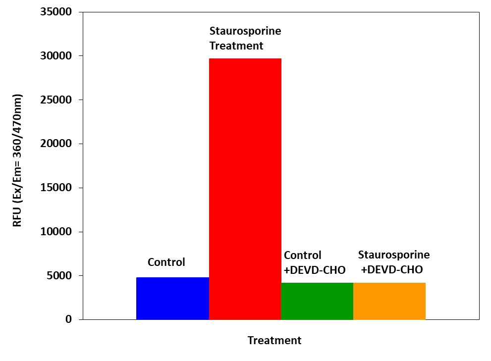
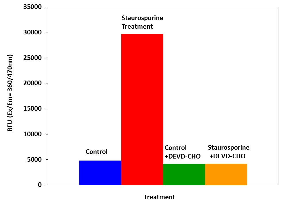
图1.使用Cell Meter Caspase 3/7活性凋亡测定试剂盒检测Jurkat细胞中的Caspase 3/7活性。 当天,将Jurkat细胞以80,000个细胞/孔/ 90 µL的浓度接种在Costar黑色96孔板中。 在有和没有1 µM星形孢菌素的情况下将细胞处理4小时,并在有或没有10 µM的胱天蛋白酶抑制剂AC-DEVD-CHO的情况下处理细胞10分钟。 加入半胱天冬酶3/7测定溶液(100μL/孔),并在室温下孵育1小时。 在Ex / Em = 360 / 470nm(截止= 420nm)下测量荧光强度。

Cell Meter Caspase 3/7活性细胞凋亡检测试剂盒 蓝色荧光

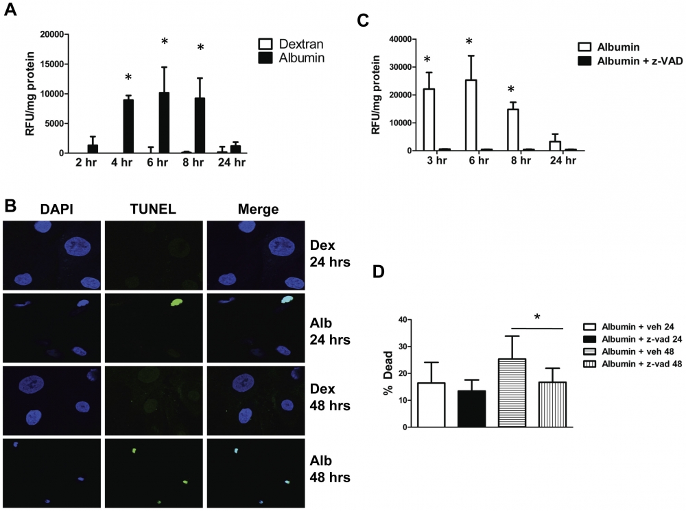
图2.白蛋白暴露上调了足细胞中的促凋亡途径。 A,活化的半胱天冬酶3/7活性相对于用5mg / ml白蛋白(实心柱)或5mg / ml右旋糖酐(实心柱)处理的足细胞中的总细胞蛋白标准化了指定的时间。 *表示与相应的右旋糖酐处理的对照相比,P <0.0001。 B,用5mg / ml葡聚糖(Dex)或5mg / ml白蛋白(Alb)处理培养的足细胞的TUNEL染色。核用DAPI染成蓝色。 TUNEL阳性细胞核是绿色的。 C,将半胱天冬酶3/7活性标准化为用5 mg / ml白蛋白或5 mg / ml白蛋白+50 µM z-VAD(泛半胱氨酸蛋白酶抑制剂)处理的足细胞中的总细胞蛋白。 z VAD很大程度上废除了caspase活性(*,P = 0.01对白蛋白+ z VAD)。 D,用锥虫蓝测定法测定的死细胞百分比,用单独的5 mg / ml白蛋白(空心柱)或5 mg / ml白蛋白+50 µM z-VAD(实心柱)处理24小时或5 mg / ml的足细胞单独使用白蛋白(水平阴影线)或5 mg / ml白蛋白+50 µM z-VAD 48小时(垂直阴影线)。 *表示在48小时时与白蛋白+ z-VAD相比,P = 0.001。 * Caspase活性是根据制造商的指导使用Caspase 3/7活性试剂盒(AAT Bioquest,Sunnyvale,CA)确定的。资料来源:Graph from Endocytosis of Albumin by Podocytes Elicits an Inflammatory Response and Induces Apoptotic Cell Death by Kayo Okamura, et al., PLoS ONE, Jan. 2013.


参考文献

Anti-Tumor Effects of Low Dose Zoledronate on Lung Cancer-Induced Spine Metastasis
Authors: Akoury, Elie and Ramirez Garcia Luna, Ana Sofia and Ahangar, Pouyan and Gao, Xiaoya and Zolotarov, Pylyp and Weber, Michael H and Rosenzweig, Derek H
Journal: Journal of clinical medicine (2019): 1212

Reversal of obesity-driven aggressiveness of endometrial cancer by metformin
Authors: Guo, Hui and Kong, Weimin and Zhang, Lu and Han, Jianjun and Clark, Leslie H and Yin, Yajie and Fang, Ziwei and Sun, Wenchuan and Wang, Ji and ong and Gilliam, Timothy P and others
Journal: American journal of cancer research (2019): 2170

Serine/Threonine Kinase 35, a Target Gene of STAT3, Regulates the Proliferation and Apoptosis of Osteosarcoma Cells
Authors: Wu, Zhong and Liu, Jie and Hu, Siyuan and Zhu, Yuchang and Li, Shaohua
Journal: Cellular Physiology and Biochemistry (2018): 808–818

Angiopoietins Modulate Survival, Migration, and the Components of the Ang-Tie2 Pathway of Chronic Lymphocytic Leukaemia (CLL) Cells In Vitro
Authors: Palma, Luis Mario Aguirre and Flamme, Hanna and Gerke, Iris and Kreuzer, Karl-Anton
Journal: Cancer Microenvironment (2016): 13–26

Hypoxia regulates TRAIL sensitivity of colorectal cancer cells through mitochondrial autophagy.
Authors: Knoll, Gertrud and Bittner, Sebastian and Kurz, Maria and Jantsch, Jonathan and Ehrenschwender, Martin
Journal: Oncotarget (2016)

Promotion of osteointegration under diabetic conditions by tantalum coating-based surface modification on 3-dimensional printed porous titanium implants
Authors: Wang, Lin and Hu, Xiaofan and Ma, Xiangyu and Ma, Zhensheng and Zhang, Yang and Lu, Yizhao and Li, Xiang and Lei, Wei and Feng, Yafei
Journal: Colloids and Surfaces B: Biointerfaces (2016): 440–452

Role of delta-like ligand-4 in chemoresistance against docetaxel in MCF-7 cells
Authors: Wang, Q and Shi, Y and Butler, HJ and Xue, J and Wang, G and Duan, P and Zheng, H
Journal: Human & Experimental Toxicology (2016): 0960327116650006

microRNA-186 inhibits cell proliferation and induces apoptosis in human esophageal squamous cell carcinoma by targeting SKP2
Authors: He, Wei and Feng, Jianfang and Zhang, Yan and Wang, Yuanyuan and Zang, Wenqiao and Zhao, Guoqiang
Journal: Laboratory Investigation (2016): 317–324

Glucose promotes cell proliferation, glucose uptake and invasion in endometrial cancer cells via AMPK/mTOR/S6 and MAPK signaling
Authors: Han, Jianjun and Zhang, Lu and Guo, Hui and Wysham, Weiya Z and Roque, Dario R and Willson, Adam K and Sheng, Xiugui and Zhou, Chunxiao and Bae-Jump, Victoria L
Journal: Gynecologic oncology (2015): 668–675

IL-7 abrogates the immunosuppressive function of human double-negative T cells by activating Akt/mTOR signaling
Authors: Allgäuer, Andrea and Schreiner, Elisabeth and Ferrazzi, Fulvia and Ekici, Arif B and Gerbitz, Armin and Mackensen, Andreas and Völkl, Simon
Journal: The Journal of Immunology (2015): 3139–3148

Cell Meter Caspase 3/7活性细胞凋亡检测试剂盒 绿色荧光-AAT Bioquest荧光染料

上海金畔生物科技有限公司代理AAT Bioquest荧光染料全线产品,欢迎访问AAT Bioquest荧光染料官网了解更多信息。
Cell Meter Caspase 3/7活性细胞凋亡检测试剂盒 绿色荧光 价格 2823
产品规格

200 Tests

产品货号

Cell Meter Caspase 3/7活性细胞凋亡检测试剂盒 绿色荧光

产品参数
Ex (nm) 500 Em (nm) 522
分子量 溶剂
存储条件
产品概述

Cell Meter Caspase 3/7活性细胞凋亡检测试剂盒是美国AAT Bioquest生产的用于检测细胞凋亡的试剂盒,Cell Meter检测试剂盒是一类用于检测细胞功能的系列工具,包括细胞活性、细胞毒性、细胞凋亡、细胞膜电位以及细胞周期等方面的指标。每种检测方案均能提供不同荧光颜色的检测方案。这些检测方案为从多角度研究细胞功能活动提供了一种十分有效的方法。Cell Meter Caspase 3/7活性细胞凋亡检测试剂盒是通过测量Caspase 3/7的活性而用于细胞凋亡检测的。因为Caspase-3 (CPP32/apopain)在细胞凋亡的起始过程中发挥着重要作用,因此其作为一个值得信赖的细胞凋亡指示剂广泛地被大家所接受。Caspase 3具有特异性的底物多肽识别序列,即天冬氨酸-谷氨酸-缬氨酸-天冬氨酸(DEVD)。本试剂盒用Z-DEVD-Rh 110-DVED-Z作为Caspase-3活性的荧光指示剂。通过Caspase 3作用,断裂Rh 110多肽,产生具有强烈绿色荧光的Rh 110,检测其在520-530 nm处的荧光强度,其激发光波长在480-500nm处。本试剂盒提供所有必需组分和佳检测方案。本检测法强大、稳定,能够用于多种荧光研究平台的高通量分析中,例如微孔板分析,免疫组化和流式细胞术。使用时96孔板时,每孔用100ul试剂,本试剂盒提供的试剂足够进行200次检测;用384微孔板时,每孔加25ul试剂,本试剂盒提供的试剂足够进行800次检测。金畔生物是AAT Bioquest的中国代理商,为您提供优质的Cell Meter Caspase 3/7活性细胞凋亡检测试剂盒。 

点击查看光谱


适用仪器


荧光酶标仪  
Ex: 490 nm
Em: 525 nm
Cutoff: 515 nm
推荐孔板: 黑色透明底板
读取模式: 顶/底读模式
实验方案

样本实验方案

简要概述

1.用测试化合物(100μL/孔/ 96孔板或25μL/孔/ 384孔板)制备细胞
2.加入等体积的Caspase 3/7工作溶液
3.在室温下孵育1小时
4.监测Ex / Em = 490/525 nm处的荧光强度(截止= 515 nm)


工作溶液配制

将50μLCaspase3/7底物(组分A)加入10mL测定缓冲液(组分B)中并充分混合以制备Caspase 3/7工作溶液。有关细胞样品制备的指南,请点击查看


操作

1.通过将10μL/孔的10X测试化合物(96孔板)或5μL/孔的5X测试化合物(384孔板)加入PBS或所需缓冲液中来处理细胞。对于空白孔(没有细胞的培养基),加入相同量的化合物缓冲液。

2.将细胞板在37℃,5%CO2,培养箱中孵育所需的一段时间(对于用喜树碱处理的Jurkat细胞,4-6小时)以诱导细胞凋亡。

3.加入100μL/孔(96孔板)或25μL/孔(384孔板)的Caspase 3/7工作溶液。

4.将板在室温下孵育至少1小时,避光。注意:如果需要,在室温下加入Caspase 3/7工作溶液10分钟后,将1μL1mM Ac-DEVD-CHO caspase 3/7抑制剂加入到选定的样品中,以确认抑制半胱天冬酶3/7样活动。

5.以800rpm离心细胞板(特别是对于非贴壁细胞)2分钟(制动关闭)。

6.用荧光酶标仪在Ex / Em = 490 / 525nm(截止= 515nm)下监测荧光强度。


数据分析

Cell Meter Caspase 3/7活性细胞凋亡检测试剂盒 绿色荧光

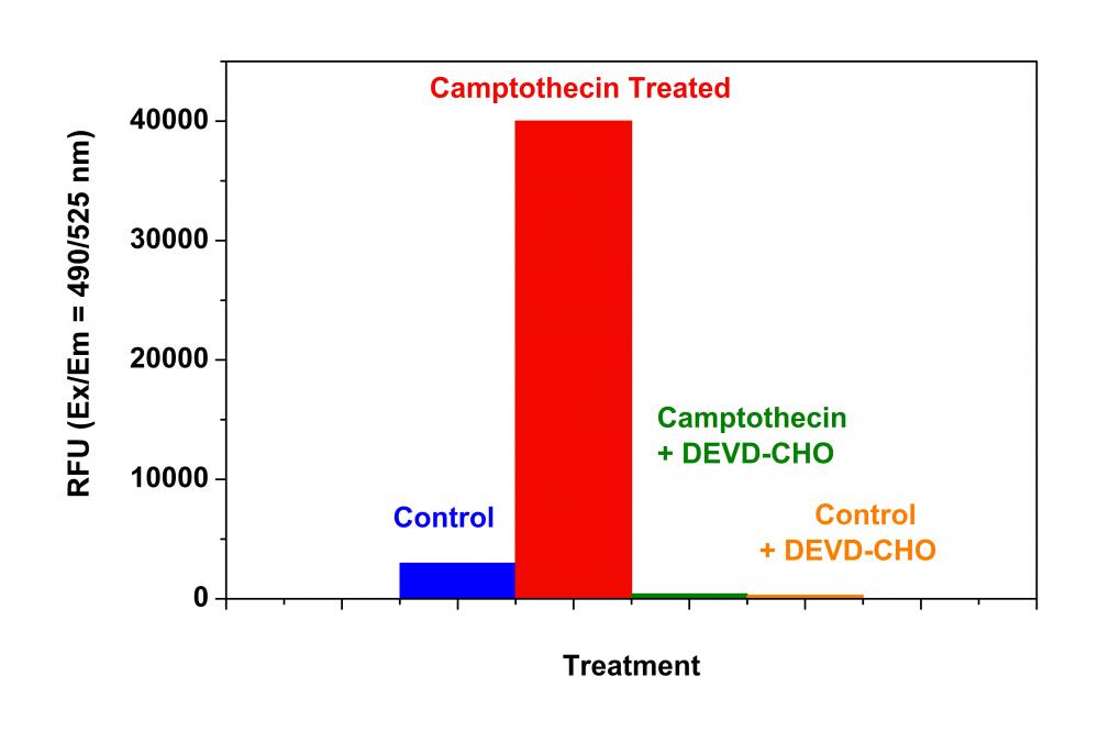
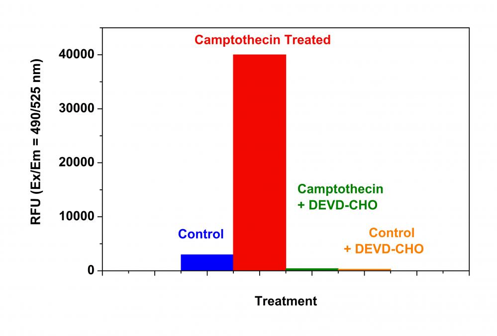
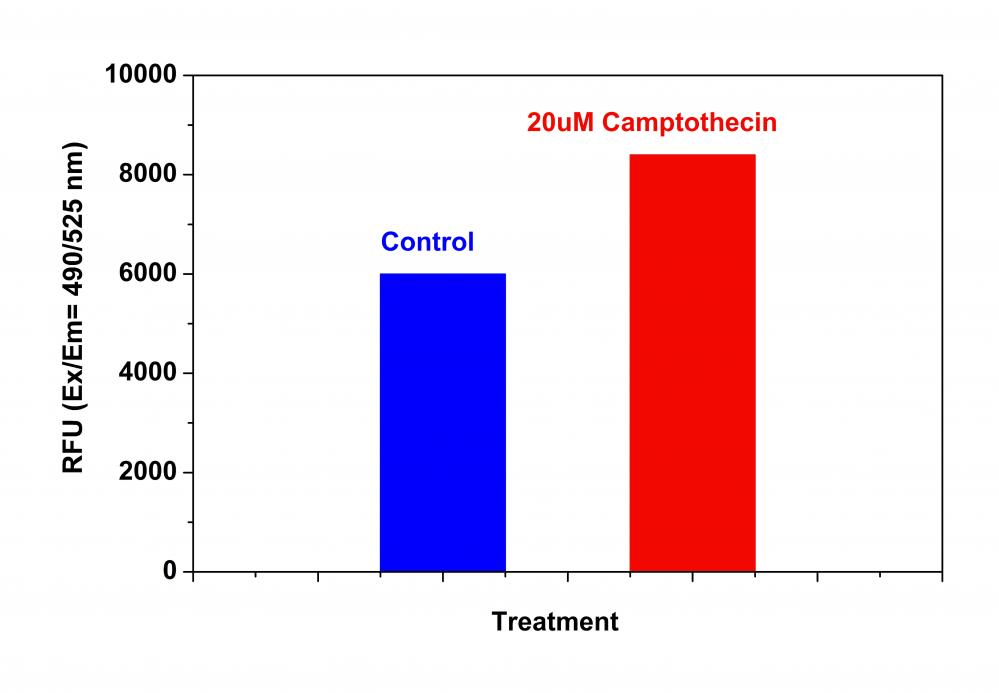
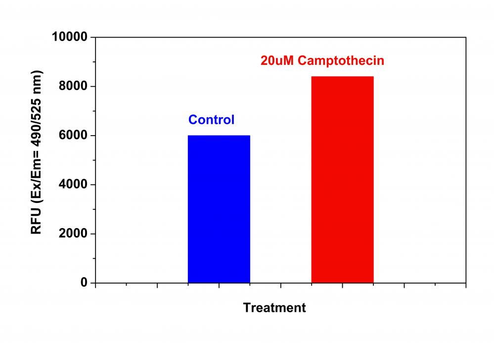
图1. Jurkat细胞中半胱天冬酶3/7活性的检测。 Jurkat细胞在以80,000个细胞/孔/90μL接种在黑色壁/透明底96孔板中。 用或不用20μM喜树碱处理细胞5小时,和/或用5μM半胱天冬酶3/7抑制剂AC-DEVD-CHO处理细胞10分钟。 加入半胱天冬酶3/7工作溶液(100μL/孔)并在室温下温育1小时。 使用NOVOstar仪器(BMG Labtech)在Ex / Em = 490 / 525nm(截止值= 515nm)下测量荧光强度。


参考文献

Hypertonicity primes malignant melanoma cells for apoptosis
Authors: Diana Nicoleta Calance, Charlotte Steixner, Stefanie Gross, Beatrice Schuler-Thurner, Gertrud Knoll, Martin Ehrenschwender
Journal: Apoptosis (2018): 1–9

Angiopoietins Modulate Survival, Migration, and the Components of the Ang-Tie2 Pathway of Chronic Lymphocytic Leukaemia (CLL) Cells In Vitro
Authors: Luis Mario Aguirre Palma, Hanna Flamme, Iris Gerke, Karl-Anton Kreuzer
Journal: Cancer Microenvironment (2016): 13–26

Hypoxia regulates TRAIL sensitivity of colorectal cancer cells through mitochondrial autophagy.
Authors: Gertrud Knoll, Sebastian Bittner, Maria Kurz, Jonathan Jantsch, Martin Ehrenschwender
Journal: Oncotarget (2016)

microRNA-186 inhibits cell proliferation and induces apoptosis in human esophageal squamous cell carcinoma by targeting SKP2
Authors: Wei He, Jianfang Feng, Yan Zhang, Yuanyuan Wang, Wenqiao Zang, Guoqiang Zhao
Journal: Laboratory Investigation (2016): 317–324

Promotion of osteointegration under diabetic conditions by tantalum coating-based surface modification on 3-dimensional printed porous titanium implants
Authors: Lin Wang, Xiaofan Hu, Xiangyu Ma, Zhensheng Ma, Yang Zhang, Yizhao Lu, Xiang Li, Wei Lei, Yafei Feng
Journal: Colloids and Surfaces B: Biointerfaces (2016): 440–452

Role of delta-like ligand-4 in chemoresistance against docetaxel in MCF-7 cells
Authors: Q Wang, Y Shi, HJ Butler, J Xue, G Wang, P Duan, H Zheng
Journal: Human & Experimental Toxicology (2016): 0960327116650006

Glucose promotes cell proliferation, glucose uptake and invasion in endometrial cancer cells via AMPK/mTOR/S6 and MAPK signaling
Authors: Jianjun Han, Lu Zhang, Hui Guo, Weiya Z Wysham, Dario R Roque, Adam K Willson, Xiugui Sheng, Chunxiao Zhou, Victoria L Bae-Jump
Journal: Gynecologic oncology (2015): 668–675

IL-7 abrogates the immunosuppressive function of human double-negative T cells by activating Akt/mTOR signaling
Authors: Andrea Allgäuer, Elisabeth Schreiner, Fulvia Ferrazzi, Arif B Ekici, Armin Gerbitz, Andreas Mackensen, Simon Völkl
Journal: The Journal of Immunology (2015): 3139–3148

Insulin improves osteogenesis of titanium implants under diabetic conditions by inhibiting reactive oxygen species overproduction via the PI3K-Akt pathway
Authors: Lin Wang, Xiong Zhao, Bo-yuan Wei, Yi Liu, Xiang-yu Ma, Jian Wang, Peng-chong Cao, Yang Zhang, Ya-bo Yan, Wei Lei
Journal: Biochimie (2015): 85–93

LGL1 modulates proliferation, apoptosis, and migration of human fetal lung fibroblasts
Authors: Hui Zhang, Neil B Sweezey, Feige Kaplan
Journal: American Journal of Physiology-Lung Cellular and Molecular Physiology (2015): L391–L402


相关产品

产品名称 货号
Cell Meter Caspase 3/7活性细胞凋亡检测试剂盒 蓝色荧光 Cat#22795
Cell Meter Caspase 3/7活性细胞凋亡检测试剂盒 红色荧光 Cat#22797
Cell Meter Caspase 3/7活性细胞凋亡检测试剂盒 绿色荧光 适合于流式细胞检测 Cat#22823

Cell Meter Caspase 3/7活性细胞凋亡检测试剂盒 红色荧光-AAT Bioquest荧光染料

上海金畔生物科技有限公司代理AAT Bioquest荧光染料全线产品,欢迎访问AAT Bioquest荧光染料官网了解更多信息。
Cell Meter Caspase 3/7活性细胞凋亡检测试剂盒 红色荧光 价格 4245
产品规格

100 Tests

产品货号

Cell Meter Caspase 3/7活性细胞凋亡检测试剂盒 红色荧光

产品参数
Ex (nm) 532 Em (nm) 619
分子量 溶剂
存储条件
产品概述

Cell Meter Caspase 3/7活性细胞凋亡检测试剂盒是美国AAT Bioquest生产的用于检测细胞凋亡的试剂盒,我们的Cell Meter 检测试剂盒是一套用于监测细胞生存力的工具。有多种参数可用于监测细胞活力。该特定试剂盒旨在通过测量Caspase 3来监测细胞凋亡。由于caspase-3(CPP32 / apopain)对于细胞凋亡的启动很重要,因此caspase 3被公认为是细胞凋亡的可靠指标。半胱天冬酶3具有对肽序列Asp-Glu-Val-Asp(DEVD)的底物选择性。该试剂盒使用Z-DEVD-ProRed 作为caspase-3活性的荧光指示剂。半胱天冬酶3对ProRed DEVD封闭肽残基的切割产生强红色荧光的ProRed ,该荧光在〜620 nm处以荧光光谱法进行监测,激发波长约为530 nm。该试剂盒可提供所有必需成分,并具有优化的测定方案,该测定适用于高通量测定。该试剂盒以96孔板每孔使用100 uL试剂,可提供足够的试剂来执行100次测定。该试剂盒以384孔板每孔使用25 uL试剂,可提供足够的试剂来执行400次测定。金畔生物是AAT Bioquest的中国代理商,为您提供优质的Cell Meter Caspase 3/7活性细胞凋亡检测试剂盒。 

点击查看光谱


适用仪器


荧光酶标仪  
Ex: 540 nm
Em: 620 nm
Cutoff: 610 nm
推荐孔板: 黑色透明底板
读取模式: 顶/底读模式
实验方案

样本实验方案

简要概述

1.用测试化合物(100μL/孔/ 96孔板或25μL/孔/ 384孔板)制备细胞
2.加入等体积的Caspase 3/7工作溶液
3.在室温下孵育1小时
4.监测Ex / Em = 540/620 nm处的荧光强度(截止= 610 nm)


溶液配制

储备溶液配制

1.Z-DEVD-ProRed 储备溶液(200X):将65 µL DMSO加到Z-DEVD-ProRed (组分A)的小瓶中,制成200X Z-DEVD-ProRed 储备液,避光储存。


工作溶液配制

将50μL的200X Z-DEVD-ProRed 储备溶液添加到10 mL的测定缓冲液(组分B)中,并充分混合以制成Caspase 3/7底物工作溶液。 注意:分装并在-20℃下存储未使用的Z-DEVD-ProRed 储备溶液(来自步骤2.2)和测定缓冲液(组分B)。 避免重复冻结/解冻循环。有关细胞样品制备的指南,请点击查看


操作步骤

1.准备细胞:

1.1 对于贴壁细胞:将细胞在生长培养基中以20,000个细胞/孔/ 90 uL接种96孔,或以5,000个细胞/孔/ 20 uL接种384孔,过夜培养。

1.2 对于非贴壁细胞:从培养基中离心细胞,然后将细胞沉淀以80,000至200,000个细胞/孔/ 90 uL悬浮在培养基中,用于96孔;或20,000至50,000个细胞/孔/ 20 uL用于384个细胞。 实验前,将板以800 rpm的速度离心2分钟。 注意:应单独评估每种细胞系,以确定诱导凋亡的佳细胞密度。

2.通过向PBS或所需的缓冲液中加入10 µL /孔的10X测试化合物(96孔板)或5 µL /孔的5X测试化合物(384孔板)来处理细胞。对于空白孔(不含细胞的培养基),添加相同量的化合物缓冲液。

3.将细胞板在37°C,5%CO2的培养箱中孵育(对于用星形孢菌素处理的Jurkat细胞为3-4小时)以诱导凋亡。

4.加入Caspase 3/7底物工作溶液(100 µL /孔/ 96孔板或25 µL /孔/ 384孔板。)

5.在避光的条件下,在室温下孵育平板至少1小时。注意:如果需要,在室温下添加Caspase 3/7工作溶液之前10分钟,向选定的样品中添加1 µL 1 mM Ac-DEVD-CHO caspase 3/7抑制剂,以确认对caspase 3 / 7-like的抑制活动。

6.使用荧光酶标仪(Ex / Em = 540/620 nm(截止= 610 nm))监控荧光强度。注意:有时,底部读取可提供更好的信噪比。如果使用底部读取模式,则以800 rpm的速度离心细胞板(特别是非粘附细胞)2分钟(制动)。


图示


Cell Meter Caspase 3/7活性细胞凋亡检测试剂盒 红色荧光

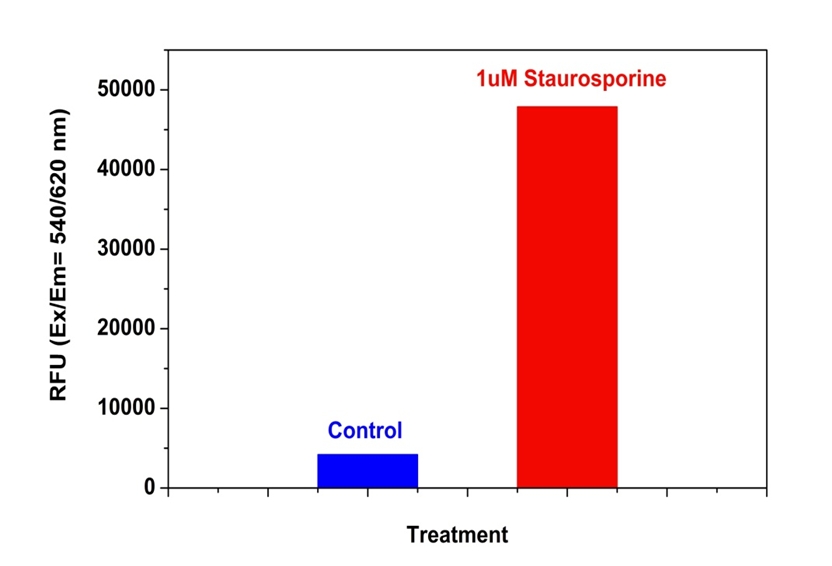
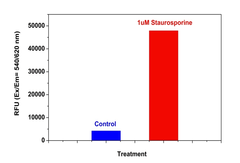
图1. Jurkat细胞中Caspase 3/7活性的检测。 当天,将Jurkat细胞以200,000个细胞/ 90 µL /孔的密度接种在Costar黑色96孔板中。 用或不用1 µM星形孢菌素处理细胞5小时。 加入半胱天冬酶3/7工作溶液(100 µL /孔)并在室温下孵育1小时。 使用FlexStation荧光酶标仪(Molecular Devices)在Ex / Em = 540/620 nm(截止= 610 nm)下测量荧光强度。


参考文献

Hypertonicity primes malignant melanoma cells for apoptosis
Authors: Diana Nicoleta Calance, Charlotte Steixner, Stefanie Gross, Beatrice Schuler-Thurner, Gertrud Knoll, Martin Ehrenschwender
Journal: Apoptosis (2018): 1–9

Angiopoietins Modulate Survival, Migration, and the Components of the Ang-Tie2 Pathway of Chronic Lymphocytic Leukaemia (CLL) Cells In Vitro
Authors: Luis Mario Aguirre Palma, Hanna Flamme, Iris Gerke, Karl-Anton Kreuzer
Journal: Cancer Microenvironment (2016): 13–26

Hypoxia regulates TRAIL sensitivity of colorectal cancer cells through mitochondrial autophagy.
Authors: Gertrud Knoll, Sebastian Bittner, Maria Kurz, Jonathan Jantsch, Martin Ehrenschwender
Journal: Oncotarget (2016)

microRNA-186 inhibits cell proliferation and induces apoptosis in human esophageal squamous cell carcinoma by targeting SKP2
Authors: Wei He, Jianfang Feng, Yan Zhang, Yuanyuan Wang, Wenqiao Zang, Guoqiang Zhao
Journal: Laboratory Investigation (2016): 317–324

Promotion of osteointegration under diabetic conditions by tantalum coating-based surface modification on 3-dimensional printed porous titanium implants
Authors: Lin Wang, Xiaofan Hu, Xiangyu Ma, Zhensheng Ma, Yang Zhang, Yizhao Lu, Xiang Li, Wei Lei, Yafei Feng
Journal: Colloids and Surfaces B: Biointerfaces (2016): 440–452

Role of delta-like ligand-4 in chemoresistance against docetaxel in MCF-7 cells
Authors: Q Wang, Y Shi, HJ Butler, J Xue, G Wang, P Duan, H Zheng
Journal: Human & Experimental Toxicology (2016): 0960327116650006

Glucose promotes cell proliferation, glucose uptake and invasion in endometrial cancer cells via AMPK/mTOR/S6 and MAPK signaling
Authors: Jianjun Han, Lu Zhang, Hui Guo, Weiya Z Wysham, Dario R Roque, Adam K Willson, Xiugui Sheng, Chunxiao Zhou, Victoria L Bae-Jump
Journal: Gynecologic oncology (2015): 668–675

IL-7 abrogates the immunosuppressive function of human double-negative T cells by activating Akt/mTOR signaling
Authors: Andrea Allgäuer, Elisabeth Schreiner, Fulvia Ferrazzi, Arif B Ekici, Armin Gerbitz, Andreas Mackensen, Simon Völkl
Journal: The Journal of Immunology (2015): 3139–3148

Insulin improves osteogenesis of titanium implants under diabetic conditions by inhibiting reactive oxygen species overproduction via the PI3K-Akt pathway
Authors: Lin Wang, Xiong Zhao, Bo-yuan Wei, Yi Liu, Xiang-yu Ma, Jian Wang, Peng-chong Cao, Yang Zhang, Ya-bo Yan, Wei Lei
Journal: Biochimie (2015): 85–93

LGL1 modulates proliferation, apoptosis, and migration of human fetal lung fibroblasts
Authors: Hui Zhang, Neil B Sweezey, Feige Kaplan
Journal: American Journal of Physiology-Lung Cellular and Molecular Physiology (2015): L391–L402


相关产品

产品名称 货号
Cell Meter Caspase 3/7活性细胞凋亡检测试剂盒 蓝色荧光 Cat#22795
Cell Meter Caspase 3/7活性细胞凋亡检测试剂盒 红色荧光 Cat#22797
Cell Meter Caspase 3/7活性细胞凋亡检测试剂盒 绿色荧光 适合于流式细胞检测 Cat#22823

Cell Meter Caspase 8活性细胞凋亡检测试剂盒 绿色荧光-AAT Bioquest荧光染料

上海金畔生物科技有限公司代理AAT Bioquest荧光染料全线产品,欢迎访问AAT Bioquest荧光染料官网了解更多信息。
Cell Meter Caspase 8活性细胞凋亡检测试剂盒 绿色荧光 价格 2823
产品规格

200 Tests

产品货号

Cell Meter Caspase 8活性细胞凋亡检测试剂盒 绿色荧光

产品参数
Ex (nm) 500 Em (nm) 522
分子量 溶剂
存储条件
产品概述

Cell Meter Caspase 8活性细胞凋亡检测试剂盒是美国AAT Bioquest生产的用于细胞凋亡的试剂盒,Cell Meter Caspase 8活性细胞凋亡检测试剂盒是通过测量Caspase 8的活性而用于细胞凋亡检测的。Caspase 8 是一种Caspase蛋白,CASP8基因所编码。在神经元变性疾病中,例如亨廷顿综合症,caspase 8扮演着重要角色。Caspase 8已被证明具有底物选择性,其肽基序为Ile-Glu-Thr-Asp (IETD)。该试剂盒使用(Ac-IETD)2-R110作为荧光指示器来检测caspas-8的活性。通过caspas-8的切割R110肽产生强绿色荧光,其发射光在520 nm和530 nm之间,激发光为480 nm和500 nm之间。这种特性使得试剂盒能适合于FITC滤光器。本试剂盒提供所有必需组分和操作流程。该实验结果稳定,快速且适应高通量筛选。它不仅能够定量凋亡细胞中caspas-8的活性还能筛选caspas-8的抑制剂。金畔生物是AAT Bioquest的中国代理商,为您提供优质的Cell Meter Caspase 8活性细胞凋亡检测试剂盒。 

点击查看光谱


适用仪器


荧光酶标仪  
Ex: 490 nm
Em: 525 nm
Cutoff: 515 nm
推荐孔板: 黑色透明底板
读取模式: 顶/底读模式
实验方案

样品实验方案

简要概述

1.用测试化合物(100μL/孔/ 96孔板或25μL/孔/ 384孔板)制备细胞
2.加入等体积的Caspase 8工作溶液(100μL/孔/ 96孔板或25μL/孔/ 384孔板)
3.在室温下孵育30分钟至1小时
4.监测Ex / Em = 490/525 nm处的荧光增加(截止= 515 nm)


工作溶液配制

将50μLCaspase8底物(组分A)加入10mL测定缓冲液(组分B)中并充分混合以制备Caspase 8工作溶液。 避光。 注意:Caspasae 8工作液不稳定,请及时使用。有关细胞样品制备的指南,请点击查看


操作步骤

1.通过将10μL/孔的10X测试化合物(96孔板)或5μL/孔的5X测试化合物(384孔板)加入PBS或所需缓冲液中来处理细胞。对于空白孔(没有细胞的培养基),加入相同量的化合物缓冲液。

2.将细胞板在5%CO 2,37℃培养箱中孵育所需的一段时间(对于用喜树碱或星形孢菌素处理的Jurkat细胞,4-6小时)以诱导细胞凋亡。

3.加入100μL/孔(96孔板)或25μL/孔(384孔板)的Caspase 8工作溶液。

4.将板在室温下孵育30分钟至1小时,避光。注意:如果需要,在室温下加入Caspase 8工作溶液10分钟前,向选定的样品中加入1μL的1 mM Ac-IETD-CHO caspase 8抑制剂,以确认对caspase 8样活性的抑制作用。

5.以800rpm离心细胞板(特别是对于非贴壁细胞)2分钟(制动关闭)。

6.用荧光酶标仪在Ex / Em = 490 / 525nm(截止= 515nm)下监测荧光增加。


数据分析

Cell Meter Caspase 8活性细胞凋亡检测试剂盒 绿色荧光

图1.使用Cell Meter Caspase 8活性细胞凋亡检测试剂盒检测Jurkat细胞中的caspase 8活性。 在Costar黑壁/透明底96孔板中以相同的以200,000个细胞/90μL/孔接种Jurkat细胞。 用或不用20μM喜树碱处理细胞4小时。 加入半胱天冬酶8工作溶液(100μL/孔)并在室温下温育1小时。 使用FlexStation 酶标仪(Molecular Devices)在Ex / Em = 490 / 525nm(截止值= 515nm)下测量荧光强度。


参考文献

Angiopoietins Modulate Survival, Migration, and the Components of the Ang-Tie2 Pathway of Chronic Lymphocytic Leukaemia (CLL) Cells In Vitro
Authors: Luis Mario Aguirre Palma, Hanna Flamme, Iris Gerke, Karl-Anton Kreuzer
Journal: Cancer Microenvironment (2016): 13–26

Hypoxia regulates TRAIL sensitivity of colorectal cancer cells through mitochondrial autophagy.
Authors: Gertrud Knoll, Sebastian Bittner, Maria Kurz, Jonathan Jantsch, Martin Ehrenschwender
Journal: Oncotarget (2016)

microRNA-186 inhibits cell proliferation and induces apoptosis in human esophageal squamous cell carcinoma by targeting SKP2
Authors: Wei He, Jianfang Feng, Yan Zhang, Yuanyuan Wang, Wenqiao Zang, Guoqiang Zhao
Journal: Laboratory Investigation (2016): 317–324

Promotion of osteointegration under diabetic conditions by tantalum coating-based surface modification on 3-dimensional printed porous titanium implants
Authors: Lin Wang, Xiaofan Hu, Xiangyu Ma, Zhensheng Ma, Yang Zhang, Yizhao Lu, Xiang Li, Wei Lei, Yafei Feng
Journal: Colloids and Surfaces B: Biointerfaces (2016): 440–452

Role of delta-like ligand-4 in chemoresistance against docetaxel in MCF-7 cells
Authors: Q Wang, Y Shi, HJ Butler, J Xue, G Wang, P Duan, H Zheng
Journal: Human & Experimental Toxicology (2016): 0960327116650006

Glucose promotes cell proliferation, glucose uptake and invasion in endometrial cancer cells via AMPK/mTOR/S6 and MAPK signaling
Authors: Jianjun Han, Lu Zhang, Hui Guo, Weiya Z Wysham, Dario R Roque, Adam K Willson, Xiugui Sheng, Chunxiao Zhou, Victoria L Bae-Jump
Journal: Gynecologic oncology (2015): 668–675

IL-7 abrogates the immunosuppressive function of human double-negative T cells by activating Akt/mTOR signaling
Authors: Andrea Allgäuer, Elisabeth Schreiner, Fulvia Ferrazzi, Arif B Ekici, Armin Gerbitz, Andreas Mackensen, Simon Völkl
Journal: The Journal of Immunology (2015): 3139–3148

Insulin improves osteogenesis of titanium implants under diabetic conditions by inhibiting reactive oxygen species overproduction via the PI3K-Akt pathway
Authors: Lin Wang, Xiong Zhao, Bo-yuan Wei, Yi Liu, Xiang-yu Ma, Jian Wang, Peng-chong Cao, Yang Zhang, Ya-bo Yan, Wei Lei
Journal: Biochimie (2015): 85–93

LGL1 modulates proliferation, apoptosis, and migration of human fetal lung fibroblasts
Authors: Hui Zhang, Neil B Sweezey, Feige Kaplan
Journal: American Journal of Physiology-Lung Cellular and Molecular Physiology (2015): L391–L402

t-BHQ Provides Protection against Lead Neurotoxicity via Nrf2/HO-1 Pathway
Authors: Fang Ye, Xiaoyi Li, Lili Li, Jing Yuan, Jun Chen
Journal: Oxidative medicine and cellular longevity (2015)


相关产品

产品名称 货号
Cell Meter Caspase 8活性细胞凋亡检测试剂盒 蓝色荧光 Cat#22812
Cell Meter Caspase 8活性细胞凋亡检测试剂盒 红色荧光 Cat#22816
Cell Meter Caspase 9活性细胞凋亡检测试剂盒 红色荧光 Cat#22817

Cell Meter Caspase 9活性细胞凋亡检测试剂盒 绿色荧光-AAT Bioquest荧光染料

上海金畔生物科技有限公司代理AAT Bioquest荧光染料全线产品,欢迎访问AAT Bioquest荧光染料官网了解更多信息。
Cell Meter Caspase 9活性细胞凋亡检测试剂盒 绿色荧光 价格 4245
产品规格

200 Tests

产品货号

Cell Meter Caspase 9活性细胞凋亡检测试剂盒 绿色荧光

产品参数
Ex (nm) 500 Em (nm) 522
分子量 溶剂
存储条件
产品概述

Cell Meter Caspase 9活性细胞凋亡检测试剂盒是美国AAT Bioquest生产的用于细胞凋亡的试剂盒,Cell Meter检测试剂盒是一类用于检测细胞功能的系列工具,包括细胞活性、细胞毒性、细胞凋亡、细胞膜电位以及细胞周期等方面的指标。每种检测方案均能提供不同荧光颜色的检测方案。这些检测方案为从多角度研究细胞功能活动提供了一种十分有效的方法。Cell Meter Caspase 9活性细胞凋亡检测试剂盒是通过测量Caspase 9的活性而用于细胞凋亡检测的。Caspase 9是CED-3亚家族中的一员。活化的Caspase 9能切割下游caspases,例如caspase-3,-6和-7,起始caspase级联反应。在正常发育的神经系统中,活化的Caspase 9是凋亡所必需的。Caspase 9已证明具有底物的选择性,其基序为Leu-Glu-His-Asp (LEHD)。本试剂盒利用(Ac-IETD)2-R110作为荧光指示器来检测caspas-9的活性。通过caspas-9的切割R110肽产生强绿色荧光,其发射光在520 nm和530 nm之间,激发光为480 nm和500 nm·之间。本试剂盒提供所有必需组分和操作流程。该实验结果稳定,快速且适应高通量筛选。它不仅能够定量凋亡细胞中caspas-9的活性还能筛选caspas-9的抑制剂。金畔生物是AAT Bioquest的中国代理商,为您提供优质的Cell Meter Caspase 9活性细胞凋亡检测试剂盒。 

点击查看光谱


适用仪器


荧光酶标仪  
Ex: 490 nm
Em: 525 nm
Cutoff: 515 nm
推荐孔板: 黑色透明底板
读取模式: 顶/底读模式
实验方案

样品实验方案

简要概述

1.用测试化合物制备细胞
2.加入等体积的Caspase 9工作溶液
3.在室温下孵育30分钟至1小时
4.在Ex / Em = 490 / 525nm处监测荧光


工作溶液配制

将50μLCaspase9底物(组分A)加入10mL测定缓冲液(组分B)中并充分混合。


操作步骤

1.通过将10μL/孔的10X测试化合物(96孔板)或5μL/孔的5X测试化合物(384孔板)加入PBS或所需缓冲液中来处理细胞。 对于空白孔(没有细胞的培养基),加入相同量的化合物缓冲液。

2.将细胞板在5%CO 2,37℃培养箱中孵育所需的一段时间以诱导细胞凋亡。

3.加入100μL/孔(96孔板)或25μL/孔(384孔板)的半胱天冬酶9工作溶液。

4.将工作溶液板在室温下孵育至少1小时,避光。 注意:如果需要,在室温下加入测定加样溶液10分钟前,向选定的样品中加入1μL的1 mM Ac-LEHD-CHO caspase 9抑制剂,以确认对caspase 9样活性的抑制。

5.将细胞板(特别是非贴壁细胞)以800rpm离心2分钟(关闭制动器)。

6.监测Ex / Em = 490 / 525nm处的荧光强度


数据分析

Cell Meter Caspase 9活性细胞凋亡检测试剂盒 绿色荧光

图1.使用Jurkat细胞中的Cell Meter Caspase 9活性细胞凋亡检测试剂盒检测caspase 9活性。 将Jurkat细胞以200,000个细胞/90μL/孔接种在Costar黑色壁/透明底96孔板中。 用10mM腺苷处理细胞48小时,而未处理的细胞用作对照。 加入半胱天冬酶9测定加载溶液(100μL/孔)并在室温下温育1小时。 使用FlexStation 酶标仪(Molecular Devices)在Ex / Em = 490 / 525nm下测量荧光强度。


参考文献

Discovery of 1-benzoyl-3-cyanopyrrolo[1,2-a]quinolines as a new series of apoptosis inducers using a cell- and caspase-based high-throughput screening assay. 2: Structure-activity relationships of the 4-, 5-, 6-, 7- and 8-positions
Authors: Kemnitzer W, Kuemmerle J, Jiang S, Sirisoma N, Kasibhatla S, Crogan-Grundy C, Tseng B, Drewe J, Cai SX.
Journal: Bioorg Med Chem Lett (2009): 3481

Discovery of substituted N’-(2-oxoindolin-3-ylidene)benzohydrazides as new apoptosis inducers using a cell- and caspase-based HTS assay
Authors: Sirisoma N, Pervin A, Drewe J, Tseng B, Cai SX.
Journal: Bioorg Med Chem Lett (2009): 2710

Selective identification of cooperatively binding fragments in a high-throughput ligation assay enables development of a picomolar caspase-3 inhibitor
Authors: Schmidt MF, El-Dahshan A, Keller S, Rademann J.
Journal: Angew Chem Int Ed Engl (2009): 6346

A dual-step fluorescence resonance energy transfer-based quenching assay for screening of caspase-3 inhibitors
Authors: Valanne A, Malmi P, Appelblom H, Niemela P, Soukka T.
Journal: Anal Biochem (2008): 71

Discovery of (naphthalen-4-yl)(phenyl)methanones and N-methyl-N-phenylnaphthalen-1-amines as new apoptosis inducers using a cell- and caspase-based HTS assay
Authors: Jiang S, Crogan-Grundy C, Drewe J, Tseng B, Cai SX.
Journal: Bioorg Med Chem Lett (2008): 5725

Discovery of 4-aryl-4H-chromenes as a new series of apoptosis inducers using a cell- and caspase-based high throughput screening assay. 4. Structure-activity relationships of N-alkyl substituted pyrrole fused at the 7,8-positions
Authors: Kemnitzer W, Drewe J, Jiang S, Zhang H, Crogan-Grundy C, Labreque D, Bubenick M, Attardo G, Denis R, Lamothe S, Gourdeau H, Tseng B, Kasibhatla S, Cai SX.
Journal: J Med Chem (2008): 417

Liver apoptosis following normothermic ischemia-reperfusion: in vivo evaluation of caspase activity by FLIVO assay in rats
Authors: Cursio R, Colosetti P, Auberger P, Gugenheim J.
Journal: Transplant Proc (2008): 2038

Modified caspase-3 assay indicates correlation of caspase-3 activity with immunity of nonhuman primates to Yersinia pestis infection
Authors: Welkos S, Norris S, Adamovicz J.
Journal: Clin Vaccine Immunol (2008): 1134

Discovery of 4-aryl-4H-chromenes as a new series of apoptosis inducers using a cell- and caspase-based high-throughput screening assay. 3. Structure-activity relationships of fused rings at the 7,8-positions
Authors: Kemnitzer W, Drewe J, Jiang S, Zhang H, Zhao J, Crogan-Grundy C, Xu L, Lamothe S, Gourdeau H, Denis R, Tseng B, Kasibhatla S, Cai SX.
Journal: J Med Chem (2007): 2858

Discovery of 5-(4-hydroxy-6-methyl-2-oxo-2H-pyran-3-yl)-7-phenyl-(E)-2,3,6,7-tetrahydro -1,4-thiazepines as a new series of apoptosis inducers using a cell- and caspase-based HTS assay
Authors: Drewe J, Kasibhatla S, Tseng B, Shelton E, Sperandio D, Yee RM, Litvak J, Sendzik M, Spencer JR, Cai SX.
Journal: Bioorg Med Chem Lett (2007): 4987


相关产品

产品名称 货号
Cell Meter Caspase 9活性细胞凋亡检测试剂盒 红色荧光 Cat#22817
Cell Meter Caspase 9活性细胞凋亡检测试剂盒 蓝色荧光 Cat#22813
Cell Meter Caspase 8活性细胞凋亡检测试剂盒 红色荧光 Cat#22816

Cell Meter 核细胞凋亡检测试剂盒 绿色荧光-AAT Bioquest荧光染料

上海金畔生物科技有限公司代理AAT Bioquest荧光染料全线产品,欢迎访问AAT Bioquest荧光染料官网了解更多信息。
Cell Meter 核细胞凋亡检测试剂盒 绿色荧光 价格 2823
产品规格

100 Tests

产品货号

Cell Meter 核细胞凋亡检测试剂盒 绿色荧光

产品参数
Ex (nm) 503 Em (nm) 527
分子量 溶剂
存储条件
产品概述

我们的Cell Meter 检测试剂盒是一套用于检测细胞生存力的工具。有多种参数可用于检测细胞活力。该特定试剂盒旨在通过测量凋亡染色质浓缩来监测细胞凋亡。与健康细胞相比,凋亡细胞的染色质紧密结合更多数量的核染料。我们的Cell Meter 核细胞凋亡测定试剂盒为所有必需成分提供了优化的测定方法,可用于检测核浓缩细胞的凋亡。这种荧光测定法是基于使用我们专有的非荧光染料对细胞中DNA含量的检测,该染料在与DNA结合后会发出强烈的荧光。在正常细胞中,Nuclear Green 不具有细胞渗透性,但是在凋亡细胞中,质膜受损或细胞代谢受损/没有细胞代谢的细胞无法阻止染料进入细胞。染料一旦进入细胞,便会与细胞内DNA结合,产生高度荧光的复合物,从而将细胞鉴定为无活力的细胞。核绿色染色;可以使用流式细胞仪(FL1通道)或荧光显微镜(FITC滤波片组)测量DCS1。该试剂盒可与我们的其他凋亡试剂一起使用,例如我们的Cell Meter NIR线粒体膜电位检测试剂盒(Cat#22802),用于细胞存活率和凋亡的多参数研究。该试剂盒经过优化,可筛选凋亡抑制剂。金畔生物是AAT Bioquest的中国代理商,为您提供优质的Cell Meter 核细胞凋亡检测试剂盒。

点击查看光谱


适用仪器


流式细胞仪  
Ex: 488 nm
Em: 530/30  nm 
通道: FITC 通道
实验方案

样品实验方案

简要概述

  1. 用5×10⁵至1×10⁶cell/ mL的密度制备含测试化合物的细胞
  2. 将5µL 200X Nuclear Green DCS1加入1 mL细胞溶液中
  3. 将细胞在37°C,5%CO2培养箱中孵育30至60分钟
  4. 沉淀细胞并将细胞重悬于1 mL生长培养基中
  5. 使用带有FL1通道的流式细胞仪分析荧光强度


实验步骤

1.对于每个样品,在1 mL温暖的培养基或您选择的缓冲液中以5×10⁵至1×10⁶细胞/ mL的密度制备细胞。注意:应单独评估每种细胞系,以确定诱导凋亡的细胞密度。

2.用测试化合物处理细胞以诱导凋亡。

3.向处理过的细胞中加入5 µL 200X Nuclear Green DCS1(组分A)。

4.将细胞溶液在37°C,5%CO2培养箱中孵育30至60分钟。注意:对于贴壁细胞,用0.5 mM EDTA轻轻提起细胞以保持细胞贴壁,并在用Nuclear Green DCS1染料上样溶液孵育之前,用含血清的培养基洗涤细胞一次。合适的孵育时间取决于所用的单个细胞类型和细胞浓度。优化每个实验的孵育时间。

5.可选:以1000 rpm离心细胞4分钟,然后将细胞重悬于1 mL的测定缓冲液(组分B)或自备缓冲液中。

6.使用带有FL1通道的流式细胞仪(Ex / Em = 490/525 nm)检测荧光强度。


参考文献

Curcumin induces cell cycle arrest and apoptosis in human osteosarcoma (HOS) cells
Authors: Lee DS, Lee MK, Kim JH.
Journal: Anticancer Res (2009): 5039

Real-time in vivo imaging of retinal cell apoptosis after laser exposure
Authors: Schmitz-Valckenberg S, Guo L, Maass A, Cheung W, Vugler A, Moss SE, Munro PM, Fitzke FW, Cordeiro MF.
Journal: Invest Ophthalmol Vis Sci (2008): 2773

Effects of Chinese herbal recipe Weichang’an in inducing apoptosis and related gene expression in human gastric cancer grafted onto nude mice
Authors: Zhao AG, Yang JK, You SF, Li T, Zhao HL, Gu Y, Tang LD, Qiu JX.
Journal: Zhong Xi Yi Jie He Xue Bao (2007): 287

Effects of neuraminidase on apoptosis of blood lymphocytes in rats with implanted Morris tumor
Authors: Grata-Borkowska U, Steciwko A, Pokorski M, Drobnik J, Gasiorowski K, Pirogowicz I, Cieslar-Marczak E.
Journal: J Physiol Pharmacol (2007): 253

Growth arrest and apoptosis induced by quercetin is not linked to adipogenic conversion of human preadipocytes
Authors: Morikawa K, Ikeda C, Nonaka M, Suzuki I.
Journal: Metabolism (2007): 1656

Morphological study on apoptosis Hela cells induced by soyasaponins
Authors: Xiao JX, Huang GQ, Zhu CP, Ren DD, Zhang SH.
Journal: Toxicol In Vitro (2007): 820

Apoptosis induction and mitochondria alteration in human HeLa tumour cells by photoproducts of Rose Bengal acetate
Authors: Panzarini E, Tenuzzo B, Palazzo F, Chionna A, Dini L.
Journal: J Photochem Photobiol B (2006): 39

Cell-free plasma DNA: a marker for apoptosis during hemodialysis
Authors: Atamaniuk J, Ruzicka K, Stuhlmeier KM, Karimi A, Eigner M, Mueller MM.
Journal: Clin Chem (2006): 523

Cytoplasmic Ca2+ signals and cellular death by apoptosis in myocardiac H9c2 cells
Authors: Lax A, Soler F, Fern and ez-Belda F.
Journal: Biochim Biophys Acta (2006): 937

Growth inhibition and apoptosis inducing mechanisms of curcumin on human ovarian cancer cell line A2780
Authors: Zheng LD, Tong QS, Wu CH.
Journal: Chin J Integr Med (2006): 126

Cell Meter Caspase 8活性细胞凋亡检测试剂盒 蓝色荧光-AAT Bioquest荧光染料

上海金畔生物科技有限公司代理AAT Bioquest荧光染料全线产品,欢迎访问AAT Bioquest荧光染料官网了解更多信息。
Cell Meter Caspase 8活性细胞凋亡检测试剂盒 蓝色荧光 价格 2823
产品规格

200 tests

产品货号

Cell Meter Caspase 8活性细胞凋亡检测试剂盒 蓝色荧光

产品参数
Ex (nm) 341 Em (nm) 441
分子量 溶剂
存储条件
产品概述

Cell Meter Caspase 8活性细胞凋亡检测试剂盒是美国AAT Bioquest生产的用于细胞凋亡检测的试剂盒,Cell Meter检测试剂盒是一类用于检测细胞功能的系列工具,包括细胞活性、细胞毒性、细胞凋亡、细胞膜电位以及细胞周期等方面的指标。每种检测方案均能提供不同荧光颜色的检测方案。这些检测方案为从多角度研究细胞功能活动提供了一种十分有效的方法。Cell Meter Caspase 8活性细胞凋亡检测试剂盒 *蓝色荧光* 是通过测量Caspase 8的活性而用于细胞凋亡检测的。Caspase 8 是一种Caspase蛋白,CASP8基因所编码。在神经元变性疾病中,例如亨廷顿综合症,caspase 8扮演着重要角色。Caspase 8已被证明具有底物选择性,其肽基序为Ile-Glu-Thr-Asp (IETD)。该试剂盒使用(Ac-IETD)2-AMC作为荧光指示器来检测caspas-8的活性。通过caspas-8的切割AMC肽产生强蓝色荧光(eX/eM = 350/450 NM)。这种特性使得试剂盒能适合于DAPI滤光器。本试剂盒提供所有必需组分和操作流程。该实验结果稳定,快速且适应高通量筛选。它不仅能够定量凋亡细胞中caspas-8的活性还能筛选caspas-8的抑制剂。金畔生物是AAT Bioquest的中国代理商,为您提供优质的Cell Meter Caspase 8活性细胞凋亡检测试剂盒。 

点击查看光谱


适用仪器


荧光酶标仪  
Ex: 370 nm
Em: 450 nm
Cutoff: 420 nm
推荐孔板: 黑色透明底板
读取模式: 顶/底读模式
实验方案

样品实验方案

简要概述

1.用测试化合物(100μL/孔/ 96孔板或25μL/孔/ 384孔板)制备细胞
2.加入等体积的Caspase 8工作溶液(100μL/孔/ 96孔板或25μL/孔/ 384孔板)
3.在室温下孵育30-60分钟
4.监测Ex / Em = 370/450 nm处的荧光增加(截止= 420 nm)


工作溶液配制

将50μLCaspase8底物(组分A)加入10mL测定缓冲液(组分B)中并充分混合以制备Caspase 8工作溶液。 避光。 注意:Caspase 8工作液不稳定,请及时使用。有关细胞样品制备的指南,请点击查看


操作步骤

1.通过将10μL/孔的10X测试化合物(96孔板)或5μL/孔的5X测试化合物(384孔板)加入PBS或所需缓冲液中来处理细胞。对于空白孔(没有细胞的培养基),加入相同量的化合物缓冲液。

2.将细胞板在5%CO 2,37℃培养箱中孵育所需的一段时间(对于用星形孢菌素处理的Jurkat细胞4-6小时)以诱导细胞凋亡。

3.加入100μL/孔(96孔板)或25μL/孔(384孔板)的Caspase 8工作溶液。

4.将Caspase 8工作溶液板在室温下孵育30至60分钟,避光。注意:如果需要,在室温下加入Caspase 8工作溶液10分钟前,向选定的样品中加入1μL的1 mM Ac-IETD-CHO caspase 8抑制剂,以确认对caspase 8样活性的抑制作用。

5.以800rpm离心细胞板(特别是对于非贴壁细胞)2分钟(制动关闭)。

6.用荧光酶标仪在Ex / Em = 370 / 450nm(截止= 420nm)下监测荧光增加。


数据分析

Cell Meter Caspase 8活性细胞凋亡检测试剂盒 蓝色荧光

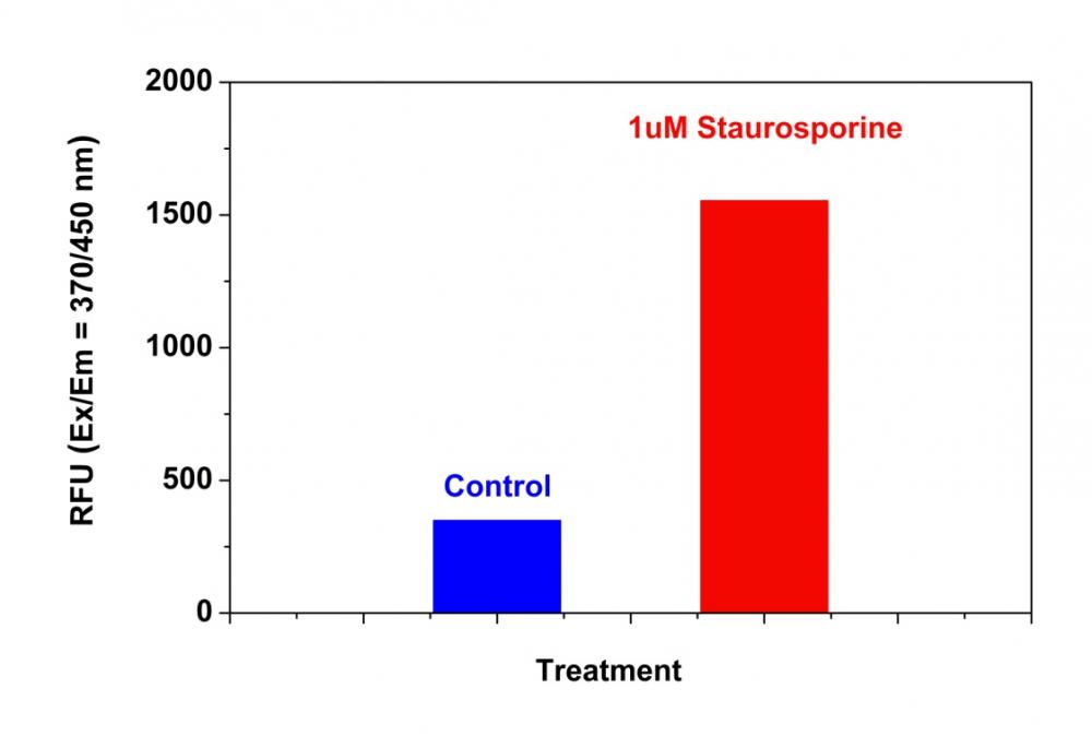
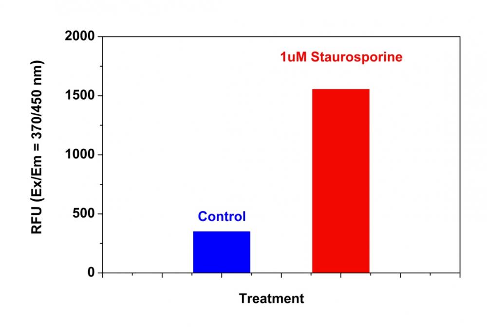
图1.使用Cell Meter Caspase 8活性细胞凋亡检测试剂盒检测Jurkat细胞中的caspase 8活性*蓝色荧光*。 在Costar黑壁/透明底96孔板中以相同的以200,000个细胞/90μL/孔接种Jurkat细胞。 用或不用1μM星形孢菌素处理细胞5小时。 加入半胱天冬酶8工作溶液(100μL/孔)并在室温下温育30分钟。 使用FlexStation 酶标仪(Molecular Devices)在Ex / Em = 370 / 450nm(截止值= 420nm)下测量荧光强度。


参考文献

Angiopoietins Modulate Survival, Migration, and the Components of the Ang-Tie2 Pathway of Chronic Lymphocytic Leukaemia (CLL) Cells In Vitro
Authors: Luis Mario Aguirre Palma, Hanna Flamme, Iris Gerke, Karl-Anton Kreuzer
Journal: Cancer Microenvironment (2016): 13–26

Hypoxia regulates TRAIL sensitivity of colorectal cancer cells through mitochondrial autophagy.
Authors: Gertrud Knoll, Sebastian Bittner, Maria Kurz, Jonathan Jantsch, Martin Ehrenschwender
Journal: Oncotarget (2016)

microRNA-186 inhibits cell proliferation and induces apoptosis in human esophageal squamous cell carcinoma by targeting SKP2
Authors: Wei He, Jianfang Feng, Yan Zhang, Yuanyuan Wang, Wenqiao Zang, Guoqiang Zhao
Journal: Laboratory Investigation (2016): 317–324

Promotion of osteointegration under diabetic conditions by tantalum coating-based surface modification on 3-dimensional printed porous titanium implants
Authors: Lin Wang, Xiaofan Hu, Xiangyu Ma, Zhensheng Ma, Yang Zhang, Yizhao Lu, Xiang Li, Wei Lei, Yafei Feng
Journal: Colloids and Surfaces B: Biointerfaces (2016): 440–452

Role of delta-like ligand-4 in chemoresistance against docetaxel in MCF-7 cells
Authors: Q Wang, Y Shi, HJ Butler, J Xue, G Wang, P Duan, H Zheng
Journal: Human & Experimental Toxicology (2016): 0960327116650006

Glucose promotes cell proliferation, glucose uptake and invasion in endometrial cancer cells via AMPK/mTOR/S6 and MAPK signaling
Authors: Jianjun Han, Lu Zhang, Hui Guo, Weiya Z Wysham, Dario R Roque, Adam K Willson, Xiugui Sheng, Chunxiao Zhou, Victoria L Bae-Jump
Journal: Gynecologic oncology (2015): 668–675

IL-7 abrogates the immunosuppressive function of human double-negative T cells by activating Akt/mTOR signaling
Authors: Andrea Allgäuer, Elisabeth Schreiner, Fulvia Ferrazzi, Arif B Ekici, Armin Gerbitz, Andreas Mackensen, Simon Völkl
Journal: The Journal of Immunology (2015): 3139–3148

Insulin improves osteogenesis of titanium implants under diabetic conditions by inhibiting reactive oxygen species overproduction via the PI3K-Akt pathway
Authors: Lin Wang, Xiong Zhao, Bo-yuan Wei, Yi Liu, Xiang-yu Ma, Jian Wang, Peng-chong Cao, Yang Zhang, Ya-bo Yan, Wei Lei
Journal: Biochimie (2015): 85–93

LGL1 modulates proliferation, apoptosis, and migration of human fetal lung fibroblasts
Authors: Hui Zhang, Neil B Sweezey, Feige Kaplan
Journal: American Journal of Physiology-Lung Cellular and Molecular Physiology (2015): L391–L402

t-BHQ Provides Protection against Lead Neurotoxicity via Nrf2/HO-1 Pathway
Authors: Fang Ye, Xiaoyi Li, Lili Li, Jing Yuan, Jun Chen
Journal: Oxidative medicine and cellular longevity (2015)


相关产品

产品名称 货号
Cell Meter Caspase 8活性细胞凋亡检测试剂盒 绿色荧光 Cat#22798
Cell Meter Caspase 8活性细胞凋亡检测试剂盒 红色荧光 Cat#22816
Cell Meter Caspase 9活性细胞凋亡检测试剂盒 红色荧光 Cat#22817

Cell Meter Caspase 9活性细胞凋亡检测试剂盒 蓝色荧光-AAT Bioquest荧光染料

上海金畔生物科技有限公司代理AAT Bioquest荧光染料全线产品,欢迎访问AAT Bioquest荧光染料官网了解更多信息。
Cell Meter Caspase 9活性细胞凋亡检测试剂盒 蓝色荧光 价格 2823
产品规格

200 Tests

产品货号

Cell Meter Caspase 9活性细胞凋亡检测试剂盒 蓝色荧光

产品参数
Ex (nm) 341 Em (nm) 441
分子量 溶剂
存储条件
产品概述

Cell Meter Caspase 9活性细胞凋亡检测试剂盒是美国AAT Bioquest生产的用于细胞凋亡检测的试剂盒,Cell Meter检测试剂盒是一类用于检测细胞功能的系列工具,包括细胞活性、细胞毒性、细胞凋亡、细胞膜电位以及细胞周期等方面的指标。每种检测方案均能提供不同荧光颜色的检测方案。这些检测方案为从多角度研究细胞功能活动提供了一种十分有效的方法。Cell Meter Caspase 9活性细胞凋亡检测试剂盒 *蓝色荧光* 是通过测量Caspase 9的活性而用于细胞凋亡检测的。Caspase 9是CED-3亚家族中的一员。活化的Caspase 9能切割下游caspases,例如caspase-3,-6和-7,起始Caspase级联反应。在正常发育的神经系统中,活化的Caspase 9是凋亡所必需的。Caspase 9具有底物的选择性,其识别底物基序为Leu-Glu-His-Asp (LEHD)。本试剂盒利用Ac-LEHD-AMC作为荧光指示器来检测caspas-9的活性。通过caspas-9的切割AMC产生强蓝色荧光,eX/eM = 350/450 nm。这种特性使得试剂盒能适合于DAPI滤光器。本试剂盒提供所有必需组分。该实验稳定、快速且适应高通量筛选的需求。它不仅能够定量凋亡细胞中caspas-9的活性还能筛选caspas-9的抑制剂。很多实验室利用本试剂盒进行高通量抑制剂的筛选。金畔生物是AAT Bioquest的中国代理商,为您提供优质的Cell Meter Caspase 9活性细胞凋亡检测试剂盒。 

点击查看光谱


适用仪器


荧光酶标仪  
Ex: 375 nm
Em: 435 nm
Cutoff: 420 nm
推荐孔板: 黑色透明底板
读取模式: 顶/底读模式
实验方案

样品实验分析

简要概述

用测试化合物(100μL/孔/ 96孔板或25μL/孔/ 384孔板)制备细胞
加入等体积的Caspase 9工作溶液(100μL/孔/ 96孔板或25μL/孔/ 384孔板)
在室温下孵育30-60分钟
监测Ex / Em = 375/435 nm处的荧光(截止= 420 nm)


工作溶液配制

将50μLCaspase9底物(组分A)加入10mL测定缓冲液(组分B)中并充分混合以制备Caspase 9工作溶液。 避光。有关细胞样品制备的指南,请点击查看

操作步骤

1.通过将10μL/孔的10X测试化合物(96孔板)或5μL/孔的5X测试化合物(384孔板)加入PBS或所需缓冲液中来处理细胞。对于空白孔(没有细胞的培养基),加入相同量的化合物缓冲液。

2.将细胞板在5%CO2,37℃培养箱中孵育所需的一段时间(对于用星形孢菌素处理的Jurkat细胞,4-6小时)以诱导细胞凋亡。

3.加入100μL/孔(96孔板)或25μL/孔(384孔板)的Caspase 9工作溶液。

4.将Caspase 9工作溶液板在室温下孵育至少1小时,避光。注意:如果需要,在室温下加入Caspase 9工作溶液前10分钟,向选定的样品中加入1μL的1 mM Ac-LEHD-CHO caspase 9抑制剂,以确认对胱天蛋白酶9样活性的抑制作用。

5.将细胞板(特别是非贴壁细胞)以800rpm离心2分钟。

6.用荧光酶标仪在Ex / Em = 375 / 435nm(截止= 420nm)下监测荧光强度。


数据分析

Cell Meter Caspase 9活性细胞凋亡检测试剂盒 蓝色荧光

图1.使用Cell Meter Caspase 9活性细胞凋亡检测试剂盒检测Jurkat细胞中的Caspase 9活性 蓝色荧光 。 将Jurkat细胞以300,000个细胞/90μL/孔接种在Costar黑色壁/透明底96孔板中。 用或不用1μM星形孢菌素处理细胞4小时。 加入半胱天冬酶9工作溶液(100μL/孔)并在室温下温育1小时。 使用FlexStation 酶标仪(Molecular Devices)在Ex / Em = 375 / 435nm(截止值= 420nm)下测量荧光强度。


参考文献

Angiopoietins Modulate Survival, Migration, and the Components of the Ang-Tie2 Pathway of Chronic Lymphocytic Leukaemia (CLL) Cells In Vitro
Authors: Luis Mario Aguirre Palma, Hanna Flamme, Iris Gerke, Karl-Anton Kreuzer
Journal: Cancer Microenvironment (2016): 13–26

Hypoxia regulates TRAIL sensitivity of colorectal cancer cells through mitochondrial autophagy.
Authors: Gertrud Knoll, Sebastian Bittner, Maria Kurz, Jonathan Jantsch, Martin Ehrenschwender
Journal: Oncotarget (2016)

microRNA-186 inhibits cell proliferation and induces apoptosis in human esophageal squamous cell carcinoma by targeting SKP2
Authors: Wei He, Jianfang Feng, Yan Zhang, Yuanyuan Wang, Wenqiao Zang, Guoqiang Zhao
Journal: Laboratory Investigation (2016): 317–324

Promotion of osteointegration under diabetic conditions by tantalum coating-based surface modification on 3-dimensional printed porous titanium implants
Authors: Lin Wang, Xiaofan Hu, Xiangyu Ma, Zhensheng Ma, Yang Zhang, Yizhao Lu, Xiang Li, Wei Lei, Yafei Feng
Journal: Colloids and Surfaces B: Biointerfaces (2016): 440–452

Role of delta-like ligand-4 in chemoresistance against docetaxel in MCF-7 cells
Authors: Q Wang, Y Shi, HJ Butler, J Xue, G Wang, P Duan, H Zheng
Journal: Human & Experimental Toxicology (2016): 0960327116650006

Glucose promotes cell proliferation, glucose uptake and invasion in endometrial cancer cells via AMPK/mTOR/S6 and MAPK signaling
Authors: Jianjun Han, Lu Zhang, Hui Guo, Weiya Z Wysham, Dario R Roque, Adam K Willson, Xiugui Sheng, Chunxiao Zhou, Victoria L Bae-Jump
Journal: Gynecologic oncology (2015): 668–675

IL-7 abrogates the immunosuppressive function of human double-negative T cells by activating Akt/mTOR signaling
Authors: Andrea Allgäuer, Elisabeth Schreiner, Fulvia Ferrazzi, Arif B Ekici, Armin Gerbitz, Andreas Mackensen, Simon Völkl
Journal: The Journal of Immunology (2015): 3139–3148

Insulin improves osteogenesis of titanium implants under diabetic conditions by inhibiting reactive oxygen species overproduction via the PI3K-Akt pathway
Authors: Lin Wang, Xiong Zhao, Bo-yuan Wei, Yi Liu, Xiang-yu Ma, Jian Wang, Peng-chong Cao, Yang Zhang, Ya-bo Yan, Wei Lei
Journal: Biochimie (2015): 85–93

LGL1 modulates proliferation, apoptosis, and migration of human fetal lung fibroblasts
Authors: Hui Zhang, Neil B Sweezey, Feige Kaplan
Journal: American Journal of Physiology-Lung Cellular and Molecular Physiology (2015): L391–L402

t-BHQ Provides Protection against Lead Neurotoxicity via Nrf2/HO-1 Pathway
Authors: Fang Ye, Xiaoyi Li, Lili Li, Jing Yuan, Jun Chen
Journal: Oxidative medicine and cellular longevity (2015)


相关产品

产品名称 货号
Cell Meter Caspase 9活性细胞凋亡检测试剂盒 绿色荧光 Cat#22799
Cell Meter Caspase 9活性细胞凋亡检测试剂盒 红色荧光 Cat#22817
Cell Meter Caspase 8活性细胞凋亡检测试剂盒 红色荧光 Cat#22816

Cell Meter Caspase 8活性细胞凋亡检测试剂盒 红色荧光-AAT Bioquest荧光染料

上海金畔生物科技有限公司代理AAT Bioquest荧光染料全线产品,欢迎访问AAT Bioquest荧光染料官网了解更多信息。
Cell Meter Caspase 8活性细胞凋亡检测试剂盒 红色荧光 价格 2823
产品规格

100 Tests

产品货号

Cell Meter Caspase 8活性细胞凋亡检测试剂盒 红色荧光

产品参数
Ex (nm) 532 Em (nm) 619
分子量 溶剂
存储条件
产品概述

Cell Meter Caspase 8活性细胞凋亡检测试剂盒是美国AAT Bioquest生产的用于细胞凋亡检测的试剂盒,Cell Meter检测试剂盒是一类用于检测细胞功能的系列工具,包括细胞活性、细胞毒性、细胞凋亡、细胞膜电位以及细胞周期等方面的指标。每种检测方案均能提供不同荧光颜色的检测方案。这些检测方案为从多角度研究细胞功能活动提供了一种十分有效的方法。Cell Meter Caspase 8活性细胞凋亡检测试剂盒 蓝色荧光 是通过测量Caspase 8的活性而用于细胞凋亡检测的。Caspase 8 是一种Caspase蛋白,CASP8基因所编码。在神经元变性疾病中,例如亨廷顿综合症,caspase 8扮演着重要角色。Caspase 8已被证明具有底物选择性,其肽基序为Ile-Glu-Thr-Asp (IETD)。该试剂盒使用(Ac-IETD)2- ProRed 作为荧光指示器来检测caspas-8的活性。通过caspas-8的切割 ProRed 肽产生强蓝色荧光(eX/eM = 530/620 NM)。这种特性使得试剂盒能适合于DAPI滤光器。本试剂盒提供所有必需组分和佳操作流程。该实验结果稳定,快速且适应高通量筛选。它不仅能够定量凋亡细胞中caspas-8的活性还能筛选caspas-8的抑制剂。金畔生物是AAT Bioquest的中国代理商,为您提供优质的Cell Meter Caspase 8活性细胞凋亡检测试剂盒。 

点击查看光谱


适用仪器


荧光酶标仪  
Ex: 450 nm
Em: 620 nm
Cutoff: 610 nm
推荐孔板: 黑色透明底板
读取模式: 顶/底读模式
实验方案

样品实验方案

简要概述

1.用测试化合物(100μL/孔/ 96孔板或25μL/孔/ 384孔板)制备细胞
2.加入等体积的Caspase 8工作溶液(100μL/孔/ 96孔板或25μL/孔/ 384孔板)
3.在室温下孵育1小时
4.在Ex / Em = 535/620 nm(截止= 610 nm)监测荧光强度(顶部或底部读取模式)


储存溶液配制

        除非另有说明,否则所有未使用的储备溶液应分成一次性等分试样,并在制备后储存在-20°C。 避免反复冻融循环。
1. Ac-IETD-ProRed 原液(200X):
将65μLDMSO加入到Ac-IETD-ProRed (组分A)的小瓶中并充分混合以制备200X Ac-IETD-ProRed 储备溶液。避光。


工作溶液配制

将50μL的200X Ac-IETD-ProRed 储备溶液加入10mL的分析缓冲液(组分B)中并充分混合以制备Caspase 8工作溶液。 避光。 注意:Caspase 8工作液不稳定,请及时使用。有关细胞样品制备的指南,请点击查看


操作步骤

1.通过将10μL/孔的10X测试化合物(96孔板)或5μL/孔的5X测试化合物(384孔板)加入PBS或所需缓冲液中来处理细胞。对于空白孔(没有细胞的培养基),加入相同量的化合物缓冲液。

2.将细胞板在37℃,5%CO2培养箱中孵育所需的一段时间(对于用星形孢菌素处理的Jurkat细胞3-4小时)以诱导细胞凋亡。

3.加入100μL/孔/ 96孔或25μL/孔/ 384孔板的Caspase 8工作溶液。

4.将板在室温下孵育至少1小时,避光。注意:如果需要,在室温下加入Caspase 8工作溶液10分钟前,向选定的样品中加入1μL的1 mM Ac-IETD-CHO caspase 8抑制剂,以确认对caspase 8样活性的抑制作用。

5.在Ex / Em = 535 / 620nm(截止= 610nm)下用荧光酶标仪(顶部或底部读取模式)监测荧光强度。注意:有时,如果使用底部读取模式,底部读数可提供更好的信号与背景比,离心细胞板(特别是非粘附细胞)在800 rpm下2分钟。


数据分析

Cell Meter Caspase 8活性细胞凋亡检测试剂盒 红色荧光

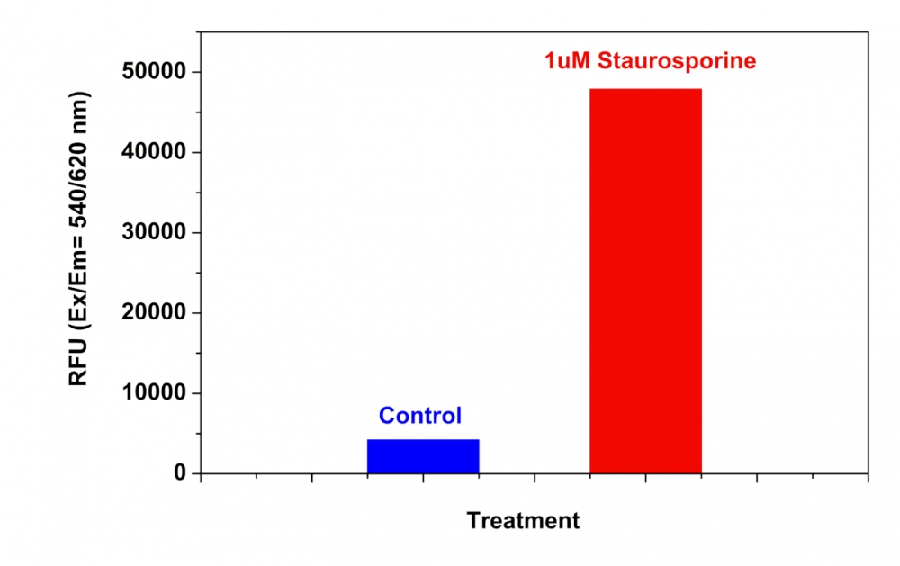
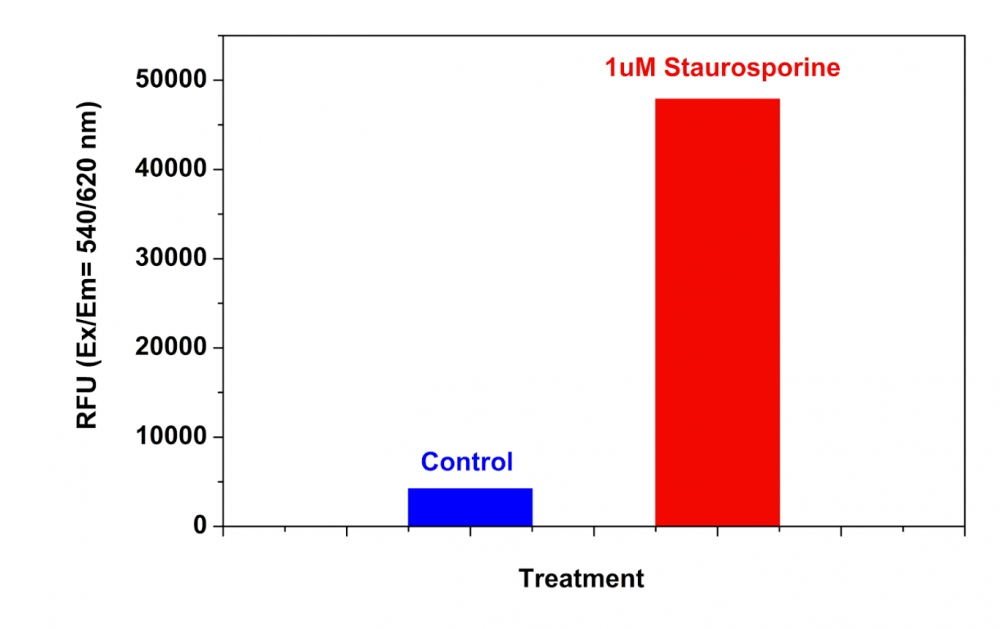
图1.使用Cell Meter Caspase 8活性细胞凋亡检测试剂盒检测Jurkat细胞中的Caspase 8活性 红色荧光 。 在Costar黑壁/透明底96孔板中以相同的以200,000个细胞/90μL/孔接种Jurkat细胞。 用或不用1μM星形孢菌素处理细胞5小时。 加入半胱天冬酶8工作溶液(100μL/孔)并在室温下温育1小时。 使用FlexStation荧光酶标仪(Molecular Devices)在Ex / Em = 540 / 620nm(截止值= 610nm)下测量荧光强度。


参考文献

Angiopoietins Modulate Survival, Migration, and the Components of the Ang-Tie2 Pathway of Chronic Lymphocytic Leukaemia (CLL) Cells In Vitro
Authors: Luis Mario Aguirre Palma, Hanna Flamme, Iris Gerke, Karl-Anton Kreuzer
Journal: Cancer Microenvironment (2016): 13–26

Hypoxia regulates TRAIL sensitivity of colorectal cancer cells through mitochondrial autophagy.
Authors: Gertrud Knoll, Sebastian Bittner, Maria Kurz, Jonathan Jantsch, Martin Ehrenschwender
Journal: Oncotarget (2016)

microRNA-186 inhibits cell proliferation and induces apoptosis in human esophageal squamous cell carcinoma by targeting SKP2
Authors: Wei He, Jianfang Feng, Yan Zhang, Yuanyuan Wang, Wenqiao Zang, Guoqiang Zhao
Journal: Laboratory Investigation (2016): 317–324

Promotion of osteointegration under diabetic conditions by tantalum coating-based surface modification on 3-dimensional printed porous titanium implants
Authors: Lin Wang, Xiaofan Hu, Xiangyu Ma, Zhensheng Ma, Yang Zhang, Yizhao Lu, Xiang Li, Wei Lei, Yafei Feng
Journal: Colloids and Surfaces B: Biointerfaces (2016): 440–452

Role of delta-like ligand-4 in chemoresistance against docetaxel in MCF-7 cells
Authors: Q Wang, Y Shi, HJ Butler, J Xue, G Wang, P Duan, H Zheng
Journal: Human & Experimental Toxicology (2016): 0960327116650006

Glucose promotes cell proliferation, glucose uptake and invasion in endometrial cancer cells via AMPK/mTOR/S6 and MAPK signaling
Authors: Jianjun Han, Lu Zhang, Hui Guo, Weiya Z Wysham, Dario R Roque, Adam K Willson, Xiugui Sheng, Chunxiao Zhou, Victoria L Bae-Jump
Journal: Gynecologic oncology (2015): 668–675

IL-7 abrogates the immunosuppressive function of human double-negative T cells by activating Akt/mTOR signaling
Authors: Andrea Allgäuer, Elisabeth Schreiner, Fulvia Ferrazzi, Arif B Ekici, Armin Gerbitz, Andreas Mackensen, Simon Völkl
Journal: The Journal of Immunology (2015): 3139–3148

Insulin improves osteogenesis of titanium implants under diabetic conditions by inhibiting reactive oxygen species overproduction via the PI3K-Akt pathway
Authors: Lin Wang, Xiong Zhao, Bo-yuan Wei, Yi Liu, Xiang-yu Ma, Jian Wang, Peng-chong Cao, Yang Zhang, Ya-bo Yan, Wei Lei
Journal: Biochimie (2015): 85–93

LGL1 modulates proliferation, apoptosis, and migration of human fetal lung fibroblasts
Authors: Hui Zhang, Neil B Sweezey, Feige Kaplan
Journal: American Journal of Physiology-Lung Cellular and Molecular Physiology (2015): L391–L402

t-BHQ Provides Protection against Lead Neurotoxicity via Nrf2/HO-1 Pathway
Authors: Fang Ye, Xiaoyi Li, Lili Li, Jing Yuan, Jun Chen
Journal: Oxidative medicine and cellular longevity (2015)


相关产品

产品名称 货号
Cell Meter Caspase 8活性细胞凋亡检测试剂盒 蓝色荧光 Cat#22812
Cell Meter Caspase 8活性细胞凋亡检测试剂盒 绿色荧光 Cat#22798
Cell Meter Caspase 9活性细胞凋亡检测试剂盒 绿色荧光 Cat#22799