核蓝 DCS1 死细胞染料-AAT Bioquest荧光染料

上海金畔生物科技有限公司代理AAT Bioquest荧光染料全线产品,欢迎访问AAT Bioquest荧光染料官网了解更多信息。
核蓝 DCS1 死细胞染料价格 1386
产品规格

0.5 ml

产品货号

核蓝 DCS1 死细胞染料

产品参数
Ex (nm) 348 Em (nm) 469
分子量 N/A 溶剂 DMSO
存储条件 在零下15度以下保存, 避免光照
产品概述

产品基本信息

产品名称:核蓝 DCS1 死细胞染料

储存条件:-20℃干燥


产品物理化学光谱特性

溶剂:DMSO

激发波长(nm):355

发射波长(nm):458


产品介绍

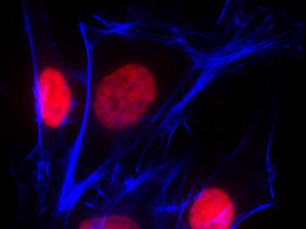
我们的Nuclear Blue DCS1是一种非荧光,DNA选择性和细胞不透性的蓝色荧光染料,用于分析死细胞中的DNA含量。 核蓝色DCS1在与双链DNA结合后具有显着增强的荧光。 它可用于荧光成像,微孔板和流式细胞仪应用。 这种DNA结合染料可用于死细胞的多色分析。

点击查看光谱

实验方案

染色细胞分析方案

注意:以下协议适用于大多数细胞类型。 生长培养基,细胞密度,其他细胞类型和因子的存在可能影响染色。 玻璃器皿上的残留洗涤剂也可能影响许多生物的染色,并导致在有或没有细胞存在的溶液中出现明亮染色的物质。

1.将Nuclear Blue DCS1(2至10μM)加入固定的,死亡或凋亡的细胞(悬浮液或粘附细胞)中,并将细胞染色15至60分钟。 在初的实验中,好尝试几种染料浓度来确定产生所需结果的佳浓度。

2.用荧光显微镜,荧光酶标仪或流式细胞仪直接分析细胞染色。 可选操作:分析前用PBS小心清洗细胞。


参考文献

One-Step Seeding of Neural Stem Cells with Vitronectin-Supplemented Medium for High-Throughput Screening Assays
Authors: Sheng Dai, Rong Li, Yan Long, Steve Titus, Jinghua Zhao, Ruili Huang, Menghang Xia, Wei Zheng
Journal: Journal of Biomolecular Screening (2016): 1112–1124

Genome shuffling of the nonconventional yeast Pichia anomala for improved sugar alcohol production
Authors: Guoqiang Zhang, Yuping Lin, Xianni Qi, Lixian Wang, Peng He, Qinhong Wang, Yanhe Ma
Journal: Microbial cell factories (2015): 1


相关产品

产品名称 货号
核绿 LCS1 活细胞染料 Cat#17540
核红 LCS1 活细胞染料 Cat#17542
核紫 LCS1 活细胞染料 Cat#17543

核绿 DCS1 死细胞染料-AAT Bioquest荧光染料

上海金畔生物科技有限公司代理AAT Bioquest荧光染料全线产品,欢迎访问AAT Bioquest荧光染料官网了解更多信息。
核绿 DCS1 死细胞染料价格 1386
产品规格

0.5 ml

产品货号

核绿 DCS1 死细胞染料

产品参数
Ex (nm) 503 Em (nm) 527
分子量 N/A 溶剂 DMSO
存储条件 在零下15度以下保存, 避免光照
产品概述

产品基本信息

产品名称:核绿 DCS1 死细胞染料

储存条件:-20℃干燥


产品物理化学光谱特性

溶剂:DMSO

激发波长(nm):503

发射波长(nm):526


产品介绍

我们的Nuclear Green DCS1是一种发荧光的,DNA选择性和不渗透细胞的染料,用于分析死亡,固定或凋亡细胞中的DNA含量。当与DNA结合后,Nuclear Green DCS1的绿色荧光显着增强。它可用于荧光成像,微孔板和流式细胞仪应用。这种具有DNA结合力的染料可用于通过适当过滤器设置对死亡,固定或凋亡细胞进行多色分析。

点击查看光谱

实验方案

染色细胞分析方案

注意:以下协议适用于大多数细胞类型。 生长培养基,细胞密度,其他细胞类型和因子的存在可能影响染色。 玻璃器皿上的残留洗涤剂也可能影响许多生物的染色,并导致在有或没有细胞存在的溶液中出现明亮染色的物质。

1.将Nuclear Green DCS1(2至10μM)加入固定的,死亡的或凋亡的细胞(悬浮液或粘附细胞)中,并将细胞染色15至60分钟。 在初的实验中,好尝试几种染料浓度来确定产生所需结果的佳浓度。 高染料浓度倾向于引起其他细胞结构的非特异性染色。

2.用荧光显微镜,荧光酶标仪或流式细胞仪直接分析细胞染色。


参考文献

One-Step Seeding of Neural Stem Cells with Vitronectin-Supplemented Medium for High-Throughput Screening Assays
Authors: Sheng Dai, Rong Li, Yan Long, Steve Titus, Jinghua Zhao, Ruili Huang, Menghang Xia, Wei Zheng
Journal: Journal of Biomolecular Screening (2016): 1112–1124

Genome shuffling of the nonconventional yeast Pichia anomala for improved sugar alcohol production
Authors: Guoqiang Zhang, Yuping Lin, Xianni Qi, Lixian Wang, Peng He, Qinhong Wang, Yanhe Ma
Journal: Microbial cell factories (2015): 1


相关产品

产品名称 货号
核绿 LCS1 活细胞染料 Cat#17540
核红 LCS1 活细胞染料 Cat#17542
核紫 LCS1 活细胞染料 Cat#17543

核橙 DCS1 死细胞染料-AAT Bioquest荧光染料

上海金畔生物科技有限公司代理AAT Bioquest荧光染料全线产品,欢迎访问AAT Bioquest荧光染料官网了解更多信息。
核橙 DCS1 死细胞染料价格 1386
产品规格

0.5 ml

产品货号

核橙 DCS1 死细胞染料

产品参数
Ex (nm) 514 Em (nm) 556
分子量 N/A 溶剂 DMSO
存储条件 在零下15度以下保存, 避免光照
产品概述

产品基本信息

产品名称:核橙 DCS1 死细胞染料

储存条件:-20℃干燥


产品物理化学光谱特性

溶剂:DMSO

激发波长(nm):528

发射波长(nm):576


产品介绍

我们的Nuclear Orange DCS1是一种荧光染料,DNA选择性和不渗透细胞的染料,可用于分析死亡,固定或凋亡细胞中的DNA含量。在与DNA结合后,Nuclear Orange DCS1的橙色荧光显着增强。它可用于荧光成像,微孔板和流式细胞仪应用。这种具有DNA结合力的染料可用于通过适当过滤器设置对死亡,固定或凋亡细胞进行多色分析。

点击查看光谱

实验方案

染色细胞分析方案

注意:以下协议适用于大多数细胞类型。 生长培养基,细胞密度,其他细胞类型和因子的存在可能影响染色。 玻璃器皿上的残留洗涤剂也可能影响许多生物的染色,并导致在有或没有细胞存在的溶液中出现明亮染色的物质。

1.将Nuclear Green DCS1(2至10μM)加入固定的,死亡的或凋亡的细胞(悬浮液或粘附细胞)中,并将细胞染色15至60分钟。 在初的实验中,好尝试几种染料浓度来确定产生所需结果的佳浓度。 高染料浓度倾向于引起其他细胞结构的非特异性染色。

2.用荧光显微镜,荧光酶标仪或流式细胞仪直接分析细胞染色。


参考文献

One-Step Seeding of Neural Stem Cells with Vitronectin-Supplemented Medium for High-Throughput Screening Assays
Authors: Sheng Dai, Rong Li, Yan Long, Steve Titus, Jinghua Zhao, Ruili Huang, Menghang Xia, Wei Zheng
Journal: Journal of Biomolecular Screening (2016): 1112–1124

Genome shuffling of the nonconventional yeast Pichia anomala for improved sugar alcohol production
Authors: Guoqiang Zhang, Yuping Lin, Xianni Qi, Lixian Wang, Peng He, Qinhong Wang, Yanhe Ma
Journal: Microbial cell factories (2015): 1


相关产品

产品名称 货号
核绿 LCS1 活细胞染料 Cat#17540
核红 LCS1 活细胞染料 Cat#17542
核紫 LCS1 活细胞染料 Cat#17543

核红 DCS1 死细胞染料-AAT Bioquest荧光染料

上海金畔生物科技有限公司代理AAT Bioquest荧光染料全线产品,欢迎访问AAT Bioquest荧光染料官网了解更多信息。
核红 DCS1 死细胞染料价格 1386
产品规格

0.5 ml

产品货号

核红 DCS1 死细胞染料

产品参数
Ex (nm) 631 Em (nm) 651
分子量 671.42 溶剂 DMSO
存储条件 在零下15度以下保存, 避免光照
产品概述

产品基本信息

产品名称:核红 DCS1 死细胞染料

储存条件:-20℃干燥


产品物理化学光谱特性

溶剂:DMSO

激发波长(nm):642

发射波长(nm):660


产品介绍

我们的Nuclear Red DCS1是一种发荧光的,DNA选择性和不渗透细胞的染料,用于分析死细胞,固定细胞或凋亡细胞中的DNA含量。结合DNA时,Nuclear Red DCS1的红色荧光显着增强。它可用于荧光成像,微孔板和流式细胞仪应用。这种具有DNA结合力的染料可用于通过适当过滤器设置对死亡,固定或凋亡细胞进行多色分析。

点击查看光谱

实验方案

染色细胞分析方案

注意:以下协议适用于大多数细胞类型。 生长培养基,细胞密度,其他细胞类型和因子的存在可能影响染色。 玻璃器皿上的残留洗涤剂也可能影响许多生物的染色,并导致在有或没有细胞存在的溶液中出现明亮染色的物质。

1.将Nuclear Red DCS1(2至10μM)加入固定的,死亡或凋亡的细胞(悬浮液或粘附细胞)中,并将细胞染色15至60分钟。 在初的实验中,好尝试几种染料浓度来确定产生所需结果的佳浓度。 高染料浓度倾向于引起其他细胞结构的非特异性染色。

2.用荧光显微镜,荧光酶标仪或流式细胞仪直接分析细胞染色。


试剂应用文献

Neural stem cells for disease modeling and evaluation of therapeutics for infantile (CLN1/PPT1) and late infantile (CLN2/TPP1) neuronal ceroid lipofuscinoses


参考文献

Neural stem cells for disease modeling and evaluation of therapeutics for infantile (CLN1/PPT1) and late infantile (CLN2/TPP1) neuronal ceroid lipofuscinoses
Authors: Ni Sima, Rong Li, Wei Huang, Miao Xu, Jeanette Beers, Jizhong Zou, Steven Titus, Elizabeth A Ottinger, Juan J Marugan, Xing Xie
Journal: Orphanet journal of rare diseases (2018): 54

Synthesis of the Anionic Hydroxypropyl-β-cyclodextrin: Poly (decamethylenephosphate) Polyrotaxane and Evaluation of its Cholesterol Efflux Potential in Niemann-Pick C1 Cells
Authors: Kerstin Egele, Shayak Samaddar, Nina Schneider, David H Thompson, Gerhard Wenz
Journal: Journal of Materials Chemistry B (2018)

One-Step Seeding of Neural Stem Cells with Vitronectin-Supplemented Medium for High-Throughput Screening Assays
Authors: Sheng Dai, Rong Li, Yan Long, Steve Titus, Jinghua Zhao, Ruili Huang, Menghang Xia, Wei Zheng
Journal: Journal of Biomolecular Screening (2016): 1112–1124

Genome shuffling of the nonconventional yeast Pichia anomala for improved sugar alcohol production
Authors: Guoqiang Zhang, Yuping Lin, Xianni Qi, Lixian Wang, Peng He, Qinhong Wang, Yanhe Ma
Journal: Microbial cell factories (2015): 1


相关产品

产品名称 货号
核绿 LCS1 活细胞染料 Cat#17540
核红 LCS1 活细胞染料 Cat#17542
核紫 LCS1 活细胞染料 Cat#17543

DNA检测染料 PUR-1-AAT Bioquest荧光染料

上海金畔生物科技有限公司代理AAT Bioquest荧光染料全线产品,欢迎访问AAT Bioquest荧光染料官网了解更多信息。
DNA检测染料 PUR-1 价格 1386
产品规格

1 mg

产品货号

DNA检测染料 PUR-1

产品参数
Ex (nm) Em (nm)
分子量 437.30 溶剂 DMSO
存储条件 在零下15度以下保存, 避免光照
产品概述

产品基本信息

产品名称:DNA检测染料 PUR-1

储存条件:-15℃避光干燥

保质期:12个月


产品物理化学光谱特性

分子量:437.30

溶剂:DMSO

激发波长(nm):459

发射波长(nm):478


产品介绍

在没有核酸的情况下,PUR-1基本不发荧光。在存在DNA和/或RNA的情况下,其荧光显着增强。在存在RNA的情况下,PUR-1在〜478 nm处具有荧光,荧光量子产率为〜40%。PUR-1染料在488 nm处可激发(在460 nm处有大激发),并且在存在核酸的情况下发出470至550 nm之间的荧光,在478 nm处具有大发射。该染料可与流式细胞仪结合用于分析血源性寄生虫感染。


参考文献

Activation of Mitochondrial Uncoupling Protein 4 and ATP-Sensitive Potassium Channel Cumulatively Decreases Superoxide Production in Insect Mitochondria
Authors: Slocinska M, Rosinski G, Jarmuszkiewicz W.
Journal: Protein Pept Lett (2016): 63
 
Rasagiline prevents cyclosporine A-sensitive superoxide flashes induced by PK11195, the initial signal of mitochondrial membrane permeabilization and apoptosis
Authors: Wu Y, Shamoto-Nagai M, Maruyama W, Osawa T, Naoi M.
Journal: J Neural Transm (Vienna) (2016): 491
 
Mitochondrial disease-related mutations at the cytochrome b-iron-sulfur protein (ISP) interface: Molecular effects on the large-scale motion of ISP and superoxide generation studied in Rhodobacter capsulatus cytochrome bc1
Authors: Ekiert R, Borek A, Kuleta P, Czernek J, Osyczka A.
Journal: Biochim Biophys Acta (2016): 1102
 
Oleuropein Prevents Neuronal Death, Mitigates Mitochondrial Superoxide Production and Modulates Autophagy in a Dopaminergic Cellular Model
Authors: Achour I, Arel-Dubeau AM, Renaud J, Legr and M, Attard E, Germain M, Martinoli MG.
Journal: Int J Mol Sci (2016): 1293
 
Manganese-superoxide dismutase (Mn-SOD) overexpression is a common event in colorectal cancers with mitochondrial microsatellite instability
Authors: Govatati S, Malempati S, Saradamma B, Divyamaanasa D, Naidu BP, Bramhachari PV, Narayana N, Shivaji S, Bhanoori M, Tamanam RR, Rao PS, Nallanchakravarthula V.
Journal: Tumour Biol. (2016)
 
Production of superoxide/hydrogen peroxide by the mitochondrial 2-oxoadipate dehydrogenase complex
Authors: Goncalves RL, Bunik VI, Br and MD., undefined
Journal: Free Radic Biol Med (2016): 247
 
A Unifying Mechanism for Mitochondrial Superoxide Production during Ischemia-Reperfusion Injury
Authors: Chouchani ET, Pell VR, James AM, Work LM, Saeb-Parsy K, Frezza C, Krieg T, Murphy MP.
Journal: Cell Metab (2016): 254
 
Rotenone-stimulated superoxide release from mitochondrial complex I acutely augments L-type Ca2+ current in A7r5 aortic smooth muscle cells
Authors: Ochi R, Dhagia V, Lakhkar A, Patel D, Wolin MS, Gupte SA.
Journal: Am J Physiol Heart Circ Physiol (2016): H1118
 
Recombinant Mitochondrial Manganese Containing Superoxide Dismutase Protects Against Ochratoxin A-Induced Nephrotoxicity
Authors: Ciarcia R, Damiano S, Squillacioti C, Mirabella N, Pagnini U, Florio A, Severino L, Capasso G, Borrelli A, Mancini A, Boffo S, Romano G, Giordano A, Florio S.
Journal: J Cell Biochem (2016): 1352
 
Mitochondrial Calpain-1 Disrupts ATP Synthase and Induces Superoxide Generation in Type 1 Diabetic Hearts: A Novel Mechanism Contributing to Diabetic Cardiomyopathy
Authors: Ni R, Zheng D, Xiong S, Hill DJ, Sun T, Gardiner RB, Fan GC, Lu Y, Abel ED, Greer PA, Peng T.
Journal: Diabetes (2016): 255

核酸染料 LDS 751 CAS 181885-68-7-AAT Bioquest荧光染料

上海金畔生物科技有限公司代理AAT Bioquest荧光染料全线产品,欢迎访问AAT Bioquest荧光染料官网了解更多信息。
核酸染料 LDS 751 CAS 181885-68-7价格 5237
产品规格

25 mg

产品货号

核酸染料 LDS 751 CAS 181885-68-7

产品参数
Ex (nm) 561 Em (nm) 712
分子量 471.98 溶剂 DMSO
存储条件 在零下15度以下保存, 避免光照
产品概述

产品基本信息

产品名称:核酸染料LDS 751

CAS:181885-68-7

储存条件:-15℃避光防潮

保质期:24个月


产品物理化学光谱特性

分子量:471.98

外观:深绿色粉末

溶剂:DMSO

激发波长(nm):561

发射波长(nm):712


产品介绍

LDS 751是一种渗透性细胞核酸染料,它在dsDNA上在〜543 nm处具有峰值激发,但可以用488 nm激光很好地激发。它是DRAQ 5 的替代产品,由于其大的大波长发射波长(〜712 nm),可用于多色分析。与LDS 751的dsDNA结合后,荧光增强程度约为20倍。LDS-751不进入细胞核,并与线粒体膜结合。因此,当使用LDS 751分析活细胞和使用LDS-751探针检测细胞时需要清楚,它不可用于细胞核检测。噻唑橙(RNA染料)和LDS-751(DNA染料)组合物已被用于将网织红细胞与其他细胞群分离。 LDS 751也已用于从有核细胞中分离红细胞。

点击查看光谱


参考文献

Exciton-controlled fluorescence: application to hybridization-sensitive fluorescent DNA probe
Authors: Okamoto A, Ikeda S, Kubota T, Yuki M, Yanagisawa H.
Journal: Nucleic Acids Symp Ser (Oxf) (2009): 49

Hybridization-sensitive fluorescent probe for long-term monitoring of intracellular RNA
Authors: Kubota T, Ikeda S, Yanagisawa H, Yuki M, Okamoto A.
Journal: Bioconjug Chem (2009): 1256

Interaction of LDS-751 with the drug-binding site of P-glycoprotein: a Trp fluorescence steady-state and lifetime study
Authors: Lugo MR, Sharom FJ.
Journal: Arch Biochem Biophys (2009): 17

Flow cytometric assessment of homeostatic aging of reticulocytes in rats
Authors: Wiczling P, Krzyzanski W.
Journal: Exp Hematol (2008): 119

Comparison of sample fixation and the use of LDS-751 or anti-CD45 for leukocyte identification in mouse whole blood for flow cytometry
Authors: Maes ML, Davidson LB, McDonagh PF, Ritter LS.
Journal: J Immunol Methods (2007): 79

Determination of the formation of dark state via depleted spontaneous emission in a complex solvated molecule
Authors: Guo X, Wang S, Xia A, Su H.
Journal: J Phys Chem A (2007): 5800

Cell cycle synchronization of Cupriavidus necator by continuous phasing measured via flow cytometry
Authors: Fritsch M, Starruss J, Loesche A, Mueller S, Bley T.
Journal: Biotechnol Bioeng (2005): 635

Exciton mobility and trapping in a MALDI matrix
Authors: Setz PD, Knochenmuss R.
Journal: J Phys Chem A (2005): 4030

Interaction of LDS-751 and rhodamine 123 with P-glycoprotein: evidence for simultaneous binding of both drugs
Authors: Lugo MR, Sharom FJ.
Journal: Biochemistry (2005): 14020

Quantitative evaluation of isothiocyanates as substrates and inhibitors of P-glycoprotein
Authors: Barecki-Roach M, Wang EJ, Johnson WW.
Journal: J Pharm Pharmacol (2003): 1251

实验方案

染色细胞分析方案

注意:以下协议适用于大多数细胞类型。 生长培养基,细胞密度,其他细胞类型和因子的存在可能影响染色。 玻璃器皿上的残留洗涤剂也可能影响许多生物的染色,并导致在有或没有细胞存在的溶液中出现明亮染色的物质。

1.制备5-10 mM DMSO储备溶液。

2.向细胞(悬浮细胞或贴壁细胞)中加入1至10μM,并将细胞染色15至60分钟。 在初的实验中,好尝试几种染料浓度来确定产生所需结果的佳浓度。 高染料浓度倾向于引起其他细胞结构的非特异性染色。

3.用荧光显微镜,荧光酶标仪或流式细胞仪直接分析细胞染色。


参考文献

Exciton-controlled fluorescence: application to hybridization-sensitive fluorescent DNA probe
Authors: Okamoto A, Ikeda S, Kubota T, Yuki M, Yanagisawa H.
Journal: Nucleic Acids Symp Ser (Oxf) (2009): 49

Hybridization-sensitive fluorescent probe for long-term monitoring of intracellular RNA
Authors: Kubota T, Ikeda S, Yanagisawa H, Yuki M, Okamoto A.
Journal: Bioconjug Chem (2009): 1256

Interaction of LDS-751 with the drug-binding site of P-glycoprotein: a Trp fluorescence steady-state and lifetime study
Authors: Lugo MR, Sharom FJ.
Journal: Arch Biochem Biophys (2009): 17

Flow cytometric assessment of homeostatic aging of reticulocytes in rats
Authors: Wiczling P, Krzyzanski W.
Journal: Exp Hematol (2008): 119

Comparison of sample fixation and the use of LDS-751 or anti-CD45 for leukocyte identification in mouse whole blood for flow cytometry
Authors: Maes ML, Davidson LB, McDonagh PF, Ritter LS.
Journal: J Immunol Methods (2007): 79

Determination of the formation of dark state via depleted spontaneous emission in a complex solvated molecule
Authors: Guo X, Wang S, Xia A, Su H.
Journal: J Phys Chem A (2007): 5800

Cell cycle synchronization of Cupriavidus necator by continuous phasing measured via flow cytometry
Authors: Fritsch M, Starruss J, Loesche A, Mueller S, Bley T.
Journal: Biotechnol Bioeng (2005): 635

Exciton mobility and trapping in a MALDI matrix
Authors: Setz PD, Knochenmuss R.
Journal: J Phys Chem A (2005): 4030

Interaction of LDS-751 and rhodamine 123 with P-glycoprotein: evidence for simultaneous binding of both drugs
Authors: Lugo MR, Sharom FJ.
Journal: Biochemistry (2005): 14020

Quantitative evaluation of isothiocyanates as substrates and inhibitors of P-glycoprotein
Authors: Barecki-Roach M, Wang EJ, Johnson WW.
Journal: J Pharm Pharmacol (2003): 1251

死细胞核染料 TWO-PRO 1 相当于TO-PRO 1 CAS 157199-59-2-AAT Bioquest荧光染料

上海金畔生物科技有限公司代理AAT Bioquest荧光染料全线产品,欢迎访问AAT Bioquest荧光染料官网了解更多信息。
死细胞核染料 TWO-PRO 1 相当于TO-PRO 1 CAS 157199-59-2价格 2823
产品规格

0.2 ml

产品货号

死细胞核染料 TWO-PRO 1  相当于TO-PRO 1  CAS 157199-59-2

产品参数
Ex (nm) 515 Em (nm) 531
分子量 645.38 溶剂 DMSO
存储条件 在零下15度以下保存, 避免光照
产品概述

产品基本信息

产品名称:死细胞核染料 TWO-PRO 1  相当于TO-PRO 1 

CAS:157199-59-2

储存条件:-15℃避光干燥

保质期:12个月


产品物理化学光谱特性

分子量:645.38

激发波长(nm):515

发射波长(nm):531


产品介绍

TWO-PRO -1在化学上等同于TO-PRO®-1(TO-PRO®是Invitrogen的商标)。TWO-PRO -1是一种碳花青二聚体,具有类似于FITC的绿色荧光。它不具有细胞渗透性,可以很容易地与Cy5和罗丹明区别开来,作为核复染和死细胞指示剂。在不存在核酸的情况下它是非荧光的,并且在与DNA结合后会产生非常明亮的荧光信号。TWO-PRO -1可在培养细胞和石蜡切片中进行强而有选择性的核染色。同时使用细胞可渗透的Nuclear Red LCS1染料和不渗透细胞的TWO-PRO -1进行标记,可用于评估细胞活力。TWO-PRO -1和Nuclear Red 的消光系数都比结合DNA的碘化丙啶大。

点击查看光谱


试剂应用文献

A toolkit for the identification of NEAT1_2/paraspeckle modulators
Authors: An, Haiyan and Elvers, Karen T and Gillespie, Jason A and Jones, Kimberley and Atack, John R and Grubisha, Olivera and Shelkovnikova, Tatyana A
Journal: Nucleic Acids Research (2022): e119–e119


参考文献

Daptomycin exerts rapid bactericidal activity against Bacillus anthracis without disrupting membrane integrity
Authors: Xing YH, Wang W, Dai SQ, Liu TY, Tan JJ, Qu GL, Li YX, Ling Y, Liu G, Fu XQ, Chen HP.
Journal: Acta Pharmacol Sin (2014): 211
 
Quantification of Candida albicans by flow cytometry using TO-PRO((R))-3 iodide as a single-stain viability dye
Authors: Kerstens M, Boulet G, Pintelon I, Hellings M, Voeten L, Delputte P, Maes L, Cos P.
Journal: J Microbiol Methods (2013): 189
 
A silicon cell cycle in a bacterial model of calcium phosphate mineralogenesis
Authors: Linton KM, Tapping CR, Adams DG, Carter RD, Shore RC, Aaron JE.
Journal: Micron (2013): 419
 
Pulsed electromagnetic field affects intrinsic and endoplasmatic reticulum apoptosis induction pathways in MonoMac6 cell line culture
Authors: Kaszuba-Zwoinska J, Chorobik P, Juszczak K, Zaraska W, Thor PJ.
Journal: J Physiol Pharmacol (2012): 537
 
Determination of the drug-DNA binding modes using fluorescence-based assays
Authors: Williams AK, Dasilva SC, Bhatta A, Rawal B, Liu M, Korobkova EA.
Journal: Anal Biochem (2012): 66
 
Transient changes in neuronal cell membrane permeability after blast exposure
Authors: Arun P, Abu-Taleb R, Valiyaveettil M, Wang Y, Long JB, Nambiar MP.
Journal: Neuroreport (2012): 342
 
A comparison of TO-PRO-1 iodide and 5-CFDA-AM staining methods for assessing viability of planktonic algae with epifluorescence microscopy
Authors: Gorokhova E, Mattsson L, Sundstrom AM.
Journal: J Microbiol Methods (2012): 216
 
SOCS-3 antagonizes pro-apoptotic effects of TRAIL and resveratrol in prostate cancer cells
Authors: Horndasch M, Culig Z.
Journal: Prostate (2011): 1357
 
The fluorescent dyes TO-PRO-3 and TOTO-3 iodide allow detection of microbial cells in soil samples without interference from background fluorescence
Authors: Henneberger R, Birch D, Bergquist P, Walter M, Anitori RP.
Journal: Biotechniques (2011): 190
 
Preclinical evaluation of novel triphenylphosphonium salts with broad-spectrum activity
Authors: Millard M, Pathania D, Shabaik Y, Taheri L, Deng J, Neamati N.
Journal: PLoS One (2010)
死细胞核染料 TWO-PRO 3 相当于TO-PRO 3 CAS 157199-63-8-AAT Bioquest荧光染料

上海金畔生物科技有限公司代理AAT Bioquest荧光染料全线产品,欢迎访问AAT Bioquest荧光染料官网了解更多信息。
死细胞核染料 TWO-PRO 3 相当于TO-PRO 3 CAS 157199-63-8价格 2823
产品规格

0.2 ml

产品货号

死细胞核染料 TWO-PRO 3  相当于TO-PRO 3  CAS 157199-63-8

产品参数
Ex (nm) 642 Em (nm) 657
分子量 671.42 溶剂 DMSO
存储条件 在零下15度以下保存, 避免光照
产品概述

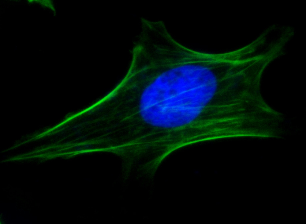
TWO-PRO -3 是一种化学成分与 TO-PRO®-3 相同的染料,后者是 Invitrogen 公司的注册商标。TWO-PRO™-3 是一种具有类似于 Cy® 5 染料的远红外荧光的碳氰化合物二聚体。它不易穿透细胞膜,因此可以清晰地与荧光素和罗丹明类染料区分开来,用作核酸对照染色和死细胞指示剂。在没有核酸的情况下它不会发光,但与 DNA 结合后会产生非常明亮的荧光信号。TWO-PRO™-3 在培养细胞和石蜡切片中提供强烈且选择性的核染色。同时使用能穿透细胞的 Nuclear Green™ LCS1 染料和不能穿透细胞的 TWO-PRO™-3 可以用来评估细胞的活性。TWO-PRO™-3 和 Nuclear Green™ 的消光系数远高于与 DNA 结合的碘化丙啶。

点击查看光谱


适用仪器


荧光显微镜  
Ex: Cy5滤波片
Em: Cy5滤波片
推荐孔板: 黑色壁/透明底


溶解方案(溶剂以说明书为准)

  0.1 mg 0.5 mg 1 mg 5 mg 10 mg
1 mM 148.938 µL 744.69 µL 1.489 mL 7.447 mL 14.894 mL
5 mM 29.788 µL 148.938 µL 297.876 µL 1.489 mL 2.979 mL
10 mM 14.894 µL 74.469 µL 148.938 µL 744.69 µL 1.489 mL
实验方案

样品实验方案

工作溶液配制

TWO-PRO -3 工作溶液

在含20 mM Hepes缓冲的Hanks缓冲液(HH缓冲液)或您选择的其他缓冲液中,制备TWO-PROTM-3的工作液。

注意:在初始实验中,最好尝试几种染料浓度以确定产生结果的最佳浓度。高染料浓度往往会导致其他细胞结构的非特异性染色。


操作步骤

注意:以下实验步骤可用于大多数细胞类型。生长基质、细胞密度、其他细胞类型及生长因子的存在可能会影响染色效果。玻璃容器上残留的洗涤剂也可能影响许多组织的染色,并会使溶液中含有或不含细胞都出现强烈荧光物质。

1.根据需要培养和处理细胞。

2.除去细胞培养基。

3.将 TWO-PRO-3 工作溶液(1 至 10 µM)添加到细胞(悬浮细胞或贴壁细胞)中,并将细胞染色 15 至 60 分钟。

4.除去染料工作溶液并添加 HH 缓冲液或您选择的缓冲液。

5.使用 Cy5 滤光片通过荧光显微镜分析细胞染色。


试剂应用文献

Synergistic Activity and Mechanism of Sanguinarine with Polymyxin B against Gram-Negative Bacterial Infections
Authors: Qiao, Luyao and Zhang, Yu and Chen, Ying and Chi, Xiangyin and Ding, Jinwen and Zhang, Hongjuan and Han, Yanxing and Zhang, Bo and Jiang, Jiandong and Lin, Yuan
Journal: Pharmaceutics (2024): 70
 
A lysosomal regulatory circuit essential for the development and function of microglia
Authors: Iyer, Harini and Shen, Kimberle and Meireles, Ana M and Talbot, William S
Journal: Science advances (2022): eabp8321


参考文献

Daptomycin exerts rapid bactericidal activity against Bacillus anthracis without disrupting membrane integrity
Authors: Xing YH, Wang W, Dai SQ, Liu TY, Tan JJ, Qu GL, Li YX, Ling Y, Liu G, Fu XQ, Chen HP.
Journal: Acta Pharmacol Sin (2014): 211
 
Quantification of Candida albicans by flow cytometry using TO-PRO((R))-3 iodide as a single-stain viability dye
Authors: Kerstens M, Boulet G, Pintelon I, Hellings M, Voeten L, Delputte P, Maes L, Cos P.
Journal: J Microbiol Methods (2013): 189
 
A silicon cell cycle in a bacterial model of calcium phosphate mineralogenesis
Authors: Linton KM, Tapping CR, Adams DG, Carter RD, Shore RC, Aaron JE.
Journal: Micron (2013): 419
 
Pulsed electromagnetic field affects intrinsic and endoplasmatic reticulum apoptosis induction pathways in MonoMac6 cell line culture
Authors: Kaszuba-Zwoinska J, Chorobik P, Juszczak K, Zaraska W, Thor PJ.
Journal: J Physiol Pharmacol (2012): 537
 
Determination of the drug-DNA binding modes using fluorescence-based assays
Authors: Williams AK, Dasilva SC, Bhatta A, Rawal B, Liu M, Korobkova EA.
Journal: Anal Biochem (2012): 66
 
Transient changes in neuronal cell membrane permeability after blast exposure
Authors: Arun P, Abu-Taleb R, Valiyaveettil M, Wang Y, Long JB, Nambiar MP.
Journal: Neuroreport (2012): 342
 
A comparison of TO-PRO-1 iodide and 5-CFDA-AM staining methods for assessing viability of planktonic algae with epifluorescence microscopy
Authors: Gorokhova E, Mattsson L, Sundstrom AM.
Journal: J Microbiol Methods (2012): 216
 
SOCS-3 antagonizes pro-apoptotic effects of TRAIL and resveratrol in prostate cancer cells
Authors: Horndasch M, Culig Z.
Journal: Prostate (2011): 1357
 
The fluorescent dyes TO-PRO-3 and TOTO-3 iodide allow detection of microbial cells in soil samples without interference from background fluorescence
Authors: Henneberger R, Birch D, Bergquist P, Walter M, Anitori RP.
Journal: Biotechniques (2011): 190
 
Preclinical evaluation of novel triphenylphosphonium salts with broad-spectrum activity
Authors: Millard M, Pathania D, Shabaik Y, Taheri L, Deng J, Neamati N.
Journal: PLoS One (2010)

死细胞核染料 DiTO -1 5 mM DMSO溶液 相当于TOTO -1 CAS 143413-84-7-AAT Bioquest荧光染料

上海金畔生物科技有限公司代理AAT Bioquest荧光染料全线产品,欢迎访问AAT Bioquest荧光染料官网了解更多信息。
死细胞核染料 DiTO -1 5 mM DMSO溶液 相当于TOTO -1 CAS 143413-84-7价格 2823
产品规格

0.2 ml

产品货号

死细胞核染料 DiTO -1  5 mM DMSO溶液 相当于TOTO -1   CAS 143413-84-7

产品参数
Ex (nm) 514 Em (nm) 531
分子量 1302.78 溶剂 Water
存储条件 在零下15度以下保存, 避免光照
产品概述

产品基本信息

产品名称:死细胞核染料 DiTO -1  5 mM DMSO溶液 相当于TOTO -1  

CAS:143413-84-7

储存条件:-15℃避光干燥

保质期:12个月


产品物理化学光谱特性

分子量:1302.78

溶剂:水

激发波长(nm):514

发射波长(nm):535


适用仪器


荧光显微镜  
激发: FITC滤波片
发射: FITC滤波片
推荐孔板: 黑色透明


产品介绍

DiTO -1在化学上等同于TOTO®-1(TOTO®是Invitrogen的商标)。DiTO -1是一种碳花青二聚体,具有类似于FITC的绿色荧光。它不具有细胞渗透性,可以很容易地与Cy5和罗丹明区别开来,作为核复染和死细胞指示剂。在不存在核酸的情况下它是非荧光的,并且在与DNA结合后会产生非常明亮的荧光信号。DiTO -1可在培养细胞和石蜡切片中进行强而有选择性的核染色。通过细胞可渗透的Nuclear Red LCS1染料和不渗透细胞的TOTO®-1进行的同时标记可用于评估细胞活力。DiTO -1和Nuclear Red 的消光系数都比结合DNA的碘化丙啶大。

点击查看光谱


参考文献

Chromatin staining of Drosophila testes
Authors: Bonaccorsi S, Giansanti MG, Cenci G, Gatti M.
Journal: Cold Spring Harb Protoc (2012)

In vivo cancer imaging by poly(ethylene glycol)-b-poly(varepsilon-caprolactone) micelles containing a near-infrared probe
Authors: Cho H, Indig GL, Weichert J, Shin HC, Kwon GS.
Journal: Nanomedicine (2012): 228

The fluorescent dyes TO-PRO-3 and TOTO-3 iodide allow detection of microbial cells in soil samples without interference from background fluorescence
Authors: Henneberger R, Birch D, Bergquist P, Walter M, Anitori RP.
Journal: Biotechniques (2011): 190

Effect of Mg ions on efficiency of gene electrotransfer and on cell electropermeabilization
Authors: Haberl S, Miklavcic D, Pavlin M.
Journal: Bioelectrochemistry (2010): 265

Visualizing nuclei in skin cryosections: viable options to 4’6-diamidino-2-phenylindol for confocal laser microscopy
Authors: Glaser K, Wilke K, Wepf R, Biel SS.
Journal: Skin Res Technol (2008): 324

Highly chlorinated Escherichia coli cannot be stained by propidium iodide
Authors: Phe MH, Dossot M, Guilloteau H, Block JC.
Journal: Can J Microbiol (2007): 664

DNA labeling in living cells
Authors: Martin RM, Leonhardt H, Cardoso MC.
Journal: Cytometry A (2005): 45

Chlorination effect on the fluorescence of nucleic acid staining dyes
Authors: Phe MH, Dossot M, Block JC.
Journal: Water Res (2004): 3729

Quinolinium, 1,1′-[1,3-propanediylbis[(dimethyliminio)-3,1-propanediyl]]bis[4-[3-(3-methyl-2(3 H)-benzothiazolylidene)-1-propen-1-yl]-,iodide (1:4)
Authors: Shan L.
Journal: In: Molecular Imaging and Contrast Agent Database (MICAD), Bethesda (MD). (2004)

Selection of fluorescent probes for flow cytometric viability assessment of Listeria monocytogenes exposed to membrane-active and oxidizing disinfectants
Authors: Luppens SB, Barbaras B, Breeuwer P, Rombouts FM, Abee T.
Journal: J Food Prot (2003): 1393

死细胞核染料 DiTO -3 相当于TO-TO 3-AAT Bioquest荧光染料

上海金畔生物科技有限公司代理AAT Bioquest荧光染料全线产品,欢迎访问AAT Bioquest荧光染料官网了解更多信息。
死细胞核染料 DiTO -3 相当于TO-TO 3 价格 2823
产品规格

0.2 ml

产品货号

死细胞核染料 DiTO -3   相当于TO-TO  3

产品参数
Ex (nm) 642 Em (nm) 661
分子量 1354.85 溶剂 Water
存储条件 在零下15度以下保存, 避免光照
产品概述

产品基本信息

货号:17576

产品名称:死细胞核染料 DiTO -3   相当于TO-TO  3  

规格:0.2ml

储存条件:-15℃避光干燥

保质期:12个月


产品物理化学光谱特性

分子量:1354.85

溶剂:水

激发波长(nm):642

发射波长(nm):660


产品介绍

DiTO -3在化学上等同于TOTO®-3(TOTO®是Invitrogen的商标)。DiTO -3是具有深红色荧光的碳花青二聚体,类似于Cy®5染料。它是不渗透细胞的,并且容易与荧光素和罗丹明区别开来,作为核复染和死细胞指示剂。在不存在核酸的情况下它是非荧光的,并且在与DNA结合后会产生非常明亮的荧光信号。DiTO -3可在培养细胞和石蜡切片中进行强而有选择性的核染色。同时使用细胞可渗透的Nuclear Green LCS1染料和不渗透细胞的DiTO®-3进行标记,可以评估细胞的生存能力。DiTO -3和Nuclear Green 的消光系数均比结合DNA的碘化丙啶大。

点击查看光谱


参考文献

Chromatin staining of Drosophila testes
Authors: Bonaccorsi S, Giansanti MG, Cenci G, Gatti M.
Journal: Cold Spring Harb Protoc (2012)

In vivo cancer imaging by poly(ethylene glycol)-b-poly(varepsilon-caprolactone) micelles containing a near-infrared probe
Authors: Cho H, Indig GL, Weichert J, Shin HC, Kwon GS.
Journal: Nanomedicine (2012): 228

The fluorescent dyes TO-PRO-3 and TOTO-3 iodide allow detection of microbial cells in soil samples without interference from background fluorescence
Authors: Henneberger R, Birch D, Bergquist P, Walter M, Anitori RP.
Journal: Biotechniques (2011): 190

Effect of Mg ions on efficiency of gene electrotransfer and on cell electropermeabilization
Authors: Haberl S, Miklavcic D, Pavlin M.
Journal: Bioelectrochemistry (2010): 265

Visualizing nuclei in skin cryosections: viable options to 4’6-diamidino-2-phenylindol for confocal laser microscopy
Authors: Glaser K, Wilke K, Wepf R, Biel SS.
Journal: Skin Res Technol (2008): 324

Highly chlorinated Escherichia coli cannot be stained by propidium iodide
Authors: Phe MH, Dossot M, Guilloteau H, Block JC.
Journal: Can J Microbiol (2007): 664

DNA labeling in living cells
Authors: Martin RM, Leonhardt H, Cardoso MC.
Journal: Cytometry A (2005): 45

Chlorination effect on the fluorescence of nucleic acid staining dyes
Authors: Phe MH, Dossot M, Block JC.
Journal: Water Res (2004): 3729

Quinolinium, 1,1′-[1,3-propanediylbis[(dimethyliminio)-3,1-propanediyl]]bis[4-[3-(3-methyl-2(3 H)-benzothiazolylidene)-1-propen-1-yl]-,iodide (1:4)
Authors: Shan L.
Journal: In: Molecular Imaging and Contrast Agent Database (MICAD), Bethesda (MD). (2004)

Selection of fluorescent probes for flow cytometric viability assessment of Listeria monocytogenes exposed to membrane-active and oxidizing disinfectants
Authors: Luppens SB, Barbaras B, Breeuwer P, Rombouts FM, Abee T.
Journal: J Food Prot (2003): 1393

死细胞核染料DiYO -1 (相当于 YOYO® 1) CAS 143413-85-8-AAT Bioquest荧光染料

上海金畔生物科技有限公司代理AAT Bioquest荧光染料全线产品,欢迎访问AAT Bioquest荧光染料官网了解更多信息。
死细胞核染料DiYO -1 (相当于 YOYO® 1) CAS 143413-85-8价格 3534
产品规格

1 mg

产品货号

死细胞核染料DiYO -1 (相当于 YOYO® 1) CAS 143413-85-8

产品参数
Ex (nm) 491 Em (nm) 508
分子量 1270.65 溶剂 DMSO
存储条件 在零下15度以下保存, 避免光照
产品概述

DiYO -1在化学上等同于YOYO®-1(YOYO®是Invitrogen的商标)。DiYO -1是一种碳花青二聚体,具有类似于FITC的绿色荧光。它不具有细胞渗透性,可以很容易地与Cy5和罗丹明区别开来,作为核复染和死细胞指示剂。在不存在核酸的情况下它是非荧光的,并且在与DNA结合后会产生非常明亮的荧光信号。DiYO -1可在培养细胞和石蜡切片中进行强而有选择性的核染色。同时使用细胞可渗透的Nuclear Red LCS1染料和不渗透细胞的DiYO®-1进行标记,可用于评估细胞活力。DiYO -1和Nuclear Red 的消光系数都比结合DNA的碘化丙啶大。

点击查看光谱

实验方案
操作步骤

1.在磷酸盐缓冲液(PBS)中将DIYO-1染料稀释至2.4 nM,然后将200 µL稀释的DIYO-1溶液直接加到冲洗过的玻片上。
2.将样品与染色剂在室温下孵育至少10-20分钟,以获得佳染色效果。
3.用水冲洗载片,以未结合的染料和PBS。 从载玻片上多余的液体。
4.加入防淬灭剂,石蜡密封。
5.用配备了标准荧光素滤光片组的荧光显微镜观察样品。
6.可以在室温或4℃的光线下保存。

 
参考文献

Correction: Photobleaching of YOYO-1 in super-resolution single DNA fluorescence imaging
Authors: J. R. Pyle
Journal: Beilstein J Nanotechnol (2018): 809-811

Ultrafast Transient Absorption Spectra of Photoexcited YOYO-1 molecules call for additional investigations of their fluorescence quenching mechanism
Authors: L. Wang
Journal: J Photochem Photobiol A Chem (2018): 411-419

Photobleaching of YOYO-1 in super-resolution single DNA fluorescence imaging
Authors: J. R. Pyle
Journal: Beilstein J Nanotechnol (2017): 2296-2306

Detection of amyloid-beta fibrils using the DNA-intercalating dye YOYO-1: Binding mode and fibril formation kinetics
Authors: D. J. Lindberg
Journal: Biochem Biophys Res Commun (2016): 313-8

Charging YOYO-1 on capillary wall for online DNA intercalation and integrating this approach with multiplex PCR and bare narrow capillary-hydrodynamic chromatography for online DNA analysis
Authors: H. Chen
Journal: Anal Chem (2015): 1518-22

Interference of ATP with the fluorescent probes YOYO-1 andYOYO-3 modifies the mechanical properties of intercalator-stained DNA confined in nanochannels
Authors: M. Roushan
Journal: Mikrochim Acta (2015): 1561-1565

Effect of YOYO-1 on the mechanical properties of DNA
Authors: B. Kundukad
Journal: Soft Matter (2014): 9721-8

Interaction of YOYO-1 with guanine-rich DNA
Authors: S. G. Datta
Journal: J Biomol Struct Dyn (2014): 1155-63

Feasibility of flow cytometry for measurements of Plasmodium falciparum parasite burden in studies in areas of malaria endemicity by use of bidimensional assessment of YOYO-1 and autofluorescence
Authors: J. J. Campo
Journal: J Clin Microbiol (2011): 968-74

Is photocleavage of DNA by YOYO-1 using a synchrotron radiation light source sequence dependent?
Authors: E. L. Gilroy
Journal: Eur Biophys J (2011): 1121-9

死细胞核染料 DiYO -1 相当于 YOYO 1 *5 mM DMSO Solution* CAS 143413-85-8-AAT Bioquest荧光染料

上海金畔生物科技有限公司代理AAT Bioquest荧光染料全线产品,欢迎访问AAT Bioquest荧光染料官网了解更多信息。
死细胞核染料 DiYO -1 相当于 YOYO 1 *5 mM DMSO Solution* CAS 143413-85-8价格 2823
产品规格

0.2 ml

产品货号

死细胞核染料 DiYO -1 相当于 YOYO  1 *5 mM DMSO Solution*  CAS 143413-85-8

产品参数
Ex (nm) 491 Em (nm) 508
分子量 1270.65 溶剂 DMSO
存储条件 在零下15度以下保存, 避免光照
产品概述

产品基本信息

产品名称:死细胞核染料DiYO -1 (相当于 YOYO® 1)

CAS:143413-85-8

储存条件:-15℃干燥

保质期:12个月


产品物理化学光谱特性

分子量:1270.65      

溶剂:水

激发波长(nm):491

发射波长(nm):509


Journal: Photochem Photobiol (2001): 585

死细胞核染料 DiYO -3 相当于YO-YO 3 CAS 156312-20-8-AAT Bioquest荧光染料

上海金畔生物科技有限公司代理AAT Bioquest荧光染料全线产品,欢迎访问AAT Bioquest荧光染料官网了解更多信息。
死细胞核染料 DiYO -3 相当于YO-YO 3 CAS 156312-20-8价格 2823
产品规格

0.2 ml

产品货号

死细胞核染料 DiYO -3   相当于YO-YO  3 CAS 156312-20-8

产品参数
Ex (nm) 612 Em (nm) 631
分子量 1322.73 溶剂 DMSO
存储条件 在零下15度以下保存, 避免光照
产品概述

产品基本信息

产品名称:死细胞核染料 DiYO -3   相当于YO-YO  3 

CAS:156312-20-8

储存条件:-15℃干燥

保质期:12个月


产品物理化学光谱特性

分子量:1322.73

溶剂:水

激发波长(nm):612

发射波长(nm):631


Journal: Photochem Photobiol (2001): 585

Cyber Green 核酸凝胶染料 [相当于 SYBR® Green] *10,000X DMSO 溶液*-AAT Bioquest荧光染料

上海金畔生物科技有限公司代理AAT Bioquest荧光染料全线产品,欢迎访问AAT Bioquest荧光染料官网了解更多信息。
Cyber Green 核酸凝胶染料 [相当于 SYBR® Green] *10,000X DMSO 溶液*价格 7092
产品规格

1 ml

产品货号

Cyber Green 核酸凝胶染料 [相当于 SYBR® Green] *10,000X DMSO 溶液*

产品参数
Ex (nm) 498 Em (nm) 522
分子量 N/A 溶剂 DMSO
存储条件 在零下15度以下保存, 避免光照
产品概述

Cyber Green 核酸凝胶染料是美国AAT Bioquest生产的一种绿色荧光核酸染料,可用于qPCR,解链曲线分析,嗜热解旋酶依赖性扩增(tHDA)的实时监测,常规溶液DNA定量和各种凝胶电泳。 DNA结合的Cyber Green 染料的激发和发射光谱非常接近荧光素(FAM)或SYBR®Green,使染料与配备488 nm氩激光的仪器或任何波长区域的可见光激发兼容。 Cyber Green 染料在热和水解方面都非常稳定,在日常处理过程中提供了便利。染料本身基本上是非荧光的,但在与dsDNA结合后变得高度荧光。荧光强度至少比溴化乙锭高一个数量级。此外,DNA / Cyber Green 复合物的荧光量子产率比DNA /溴化乙锭的荧光量子产率高5倍以上。它可以在电泳(预染色)之前,以及在电泳(后染色)之后使用,并且通过毛细管电泳分离染色DNA。它也常用于分子生物学,不含酶(例如:Taq酶,逆转录酶,内切核酸酶,T4连接酶)抑制,并且不干扰Southern印迹技术。金畔生物是AAT Bioquest的中国代理商,为您提供优质的Cyber Green 核酸凝胶染料。 

点击查看光谱

实验方案

染色方案

        我们发现通过后染色实现了大的灵敏度,这也了染料干扰DNA迁移的可能性。虽然预制方案更方便,但一些DNA样本可能会出现迁移,强烈建议凝胶时间不超过2小时。建议使用以下方案。 可能需要进行一些比较以确定哪一个更符合您的需求。


1.染色后方案

1.1根据您的标准方案运行凝胶。

1.2通过将10,000X储备试剂稀释到PH 7.5  –  8缓冲液中制备1X Cyber Green™工作溶液(例如,TAE,TBE或TE优选pH8.0)。

注意:在水中制备的染色溶液不如在缓冲液中制备的染色溶液稳定,必须在24小时内使用,以确保大的染色灵敏度。另外,在pH低于约7.5或高于8.0的缓冲液中制备的染色溶液不太稳定并且显示出降低的染色效力。

1.3将凝胶放入合适的聚丙烯容器中。轻轻加入足量的1X染色

 溶液浸没凝胶。

注意:不要使用玻璃容器,因为它会吸附染色溶液中的大部分染料。

1.4在室温下轻轻搅拌凝胶约30分钟,避光。

注意:染色溶液可以在黑暗中储存(好是冷藏)一周,多重复使用2-3次。

1.5使用254 nm透射仪或使用长路绿色滤光片(如SYBR®滤光片或GelStar®滤光片)的激光凝胶扫描仪对染色的凝胶成像。


2.预制方案

2.1使用标准方案制备琼脂糖凝胶溶液。

2.2在浇注凝胶前将10,000X Cyber Green™原液试剂以1:10,000稀释到凝胶溶液中并充分混合。

2.3根据您的标准方案运行凝胶。

2.4使用254 nm透射仪或使用长路径绿色滤光片(如SYBR®滤光片或GelStar®滤光片)的激光凝胶扫描仪对染色的凝胶成像。


3.电泳前的DNA染色

3.1在电泳前,用1:10,000稀释的染料(TE,TBE或TAE)孵育DNA至少15分钟。

3.2根据您的标准方案运行凝胶。

3.3使用254 nm透射仪或使用长路绿色滤光片(如SYBR®滤光片或GelStar®滤光片)的激光凝胶扫描仪对染色的凝胶成像。


参考文献

Plasmalemma permeability and necrotic cell death phenotypes after intracerebral hemorrhage in mice
Authors: Zhu X, Tao L, Tejima-Mandeville E, Qiu J, Park J, Garber K, Ericsson M, Lo EH, Whalen MJ.
Journal: Stroke (2012): 524

Intra-organ Biodistribution of Gold Nanoparticles Using Intrinsic Two-photon Induced Photoluminescence
Authors: Park J, Estrada A, Schwartz JA, Diagaradjane P, Krishnan S, Dunn AK, Tunnell JW.
Journal: Lasers Surg Med (2010): 630

Screening by imaging: scaling up single-DNA-molecule analysis with a novel parabolic VA-TIRF reflector and noise-reduction techniques
Authors: van ‘t Hoff M, Reuter M, Dryden DT, Oheim M.
Journal: Phys Chem Chem Phys (2009): 7713

Novel anatomic structures in the brain and spinal cord of rabbit that may belong to the Bonghan system of potential acupuncture meridians
Authors: Lee BC, Kim S, Soh KS.
Journal: J Acupunct Meridian Stud (2008): 29

Triplet fraction buildup effect of the DNA-YOYO complex studied with fluorescence correlation spectroscopy
Authors: Shimizu M, Sasaki S, Kinjo M.
Journal: Anal Biochem (2007): 87

DNA length evaluation using cyanine dye and fluorescence correlation spectroscopy
Authors: Shimizu M, Sasaki S, Tsuruoka M.
Journal: Biomacromolecules (2005): 2703

Implementation of accurate and fast DNA cytometry by confocal microscopy in 3D
Authors: Ploeger LS, Huisman A, van der Gugten J, van der Giezen DM, Belien JA, Abbaker AY, Dullens HF, Grizzle W, Poulin NM, Meijer GA, van Diest PJ.
Journal: Cell Oncol (2005): 225

Green-light transilluminator for the detection without photodamage of proteins and DNA labeled with different fluorescent dyes
Authors: Alba FJ, Bermudez A, Daban JR.
Journal: Electrophoresis (2001): 399

Oxazole yellow homodimer YOYO-1-labeled DNA: a fluorescent complex that can be used to assess structural changes in DNA following formation and cellular delivery of cationic lipid DNA complexes
Authors: Wong M, Kong S, Dragowska WH, Bally MB.
Journal: Biochim Biophys Acta (2001): 61

Photophysical properties of fluorescent DNA-dyes bound to single- and double-stranded DNA in aqueous buffered solution
Authors: Cosa G, Focsaneanu KS, McLean JR, McNamee JP, Scaiano JC.
Journal: Photochem Photobiol (2001): 585


相关产品

产品名称 货号
Cyber Orange 核酸凝胶染料 10000X DMSO Cat#17595

Cyber Orange 核酸凝胶染料 10000X DMSO-AAT Bioquest荧光染料

上海金畔生物科技有限公司代理AAT Bioquest荧光染料全线产品,欢迎访问AAT Bioquest荧光染料官网了解更多信息。
Cyber Orange 核酸凝胶染料 10000X DMSO 价格 4245
产品规格

1 ml

产品货号

Cyber Orange 核酸凝胶染料 10000X DMSO

产品参数
Ex (nm) 496 Em (nm) 539
分子量 749.48 溶剂 DMSO
存储条件 在零下15度以下保存, 避免光照
产品概述

Cyber​​ Orange 是一种出色的核酸凝胶染料,与核酸结合后显示出较大的荧光增强。它具有与SYBR®Gold相同的光谱特性,因此可以很好地替代SYBR®Gold(SYBR®Gold是Invitrogen的商标)。它是可用于检测琼脂糖和聚丙烯酰胺凝胶中的DNA的最敏感的染色剂之一。Cyber​​ Orange对DNA的敏感性高于对RNA的敏感性,非常适合与SYBR Gold仪器设置相同的激光扫描仪一起使用。Cyber​​琼脂糖对琼脂糖凝胶中的DNA比溴化乙锭敏感得多,并且在凝胶上可检测到低至皮克dsDNA。

点击查看光谱

实验方案

样品实验方案

溶液配制

工作溶液

Cyber​​ Orange 工作溶液(1X):
通过将10,000X储备试剂稀释到pH 7.5-8缓冲液(例如TAE,TBE或TE,最好是pH 8.0)中来制成1X Cyber​​ Orange 工作溶液。 注意:  在水中配制的染色溶液比在缓冲液中配制的溶液不稳定,必须在24小时内使用以确保最大的染色敏感性。 注意:  此外,在pH低于7.5或高于8.0的缓冲液中制备的染色溶液稳定性较差,并且染色效果降低。


操作步骤

1.染色后方案

1.1根据您的标准方案运行凝胶。

1.2将凝胶放在合适的聚丙烯容器中。轻轻加入足量的1X Cyber​​ Orange 工作溶液以浸没凝胶。 注意:  请勿使用玻璃容器,因为它会吸收染色溶液中的许多染料。

1.3避开光线,在室温下轻轻搅拌凝胶约30分钟。 注意:  染色溶液可以在黑暗中(最好冷藏)保存一周,并可以重复使用2-3次。

1.4使用长路径绿色滤光片(例如SYBR®滤光片或GelStar®滤光片),使用254 nm透照器或基于激光的凝胶扫描仪对染色的凝胶成像。

2.预染方案

2.1使用标准方案准备琼脂糖凝胶溶液。

2.2在凝胶中添加1X Cyber​​ Orange™工作溶液并充分混合。

2.3根据您的标准方案运行凝胶。

2.4使用长路径绿色滤光片(例如SYBR®滤光片或GelStar®滤光片),使用254 nm透照器或基于激光的凝胶扫描仪对染色的凝胶成像。

3.电泳前的DNA染色

3.1电泳前,将DNA与染料的1:1000至1:3000稀释液(在TE,TBE或TAE中)孵育至少15分钟。

3.2根据您的标准方案运行凝胶。

3.3使用254 nm透照器或使用长距离绿色滤光片(例如SYBR®滤光片或GelStar®滤光片)的基于激光的凝胶扫描仪对染色的凝胶成像。


参考文献

A Broadly Reactive One-Step SYBR Green I Real-Time RT-PCR Assay for Rapid Detection of Murine Norovirus
Authors: Hanaki K, Ike F, Kajita A, Yasuno W, Yanagiba M, Goto M, Sakai K, Ami Y, Kyuwa S.
Journal: PLoS One (2014): e98108

A SYBR Green I based real time RT-PCR assay for specific detection and quantitation of Peste des petits ruminants virus
Authors: Abera T, Thangavelu A, Joy Chandran ND, Raja A.
Journal: BMC Vet Res (2014): 22

A SYBR-green I quantitative real-time reverse transcription-PCR assay for rabies viruses with different virulence
Authors: Wang L, Liu Y, Zhang S, Wang Y, Zhao J, Miao F, Hu R.
Journal: Virol Sin (2014): 131

Comparison of SYBR Green and TaqMan methods in quantitative real-time polymerase chain reaction analysis of four adenosine receptor subtypes
Authors: Tajadini M, Panjehpour M, Javanmard SH.
Journal: Adv Biomed Res (2014): 85

Detection and characterization of Leishmania (Leishmania) and Leishmania (Viannia) by SYBR green-based real-time PCR and high resolution melt analysis targeting kinetoplast minicircle DNA
Authors: Ceccarelli M, Galluzzi L, Migliazzo A, Magnani M.
Journal: PLoS One (2014): e88845

Detection of Cardamom mosaic virus and Banana bract mosaic virus in cardamom using SYBR Green based reverse transcription-quantitative PCR
Authors: Siljo A, Bhat AI, Biju CN.
Journal: Virusdisease (2014): 137

Development and comparative evaluation of SYBR Green I-based one-step real-time RT-PCR assay for detection and quantification of West Nile virus in human patients
Authors: Kumar JS, Saxena D, Parida M.
Journal: Mol Cell Probes. (2014)

Development and evaluation of SYBR Green-I based quantitative PCR assays for herpes simplex virus type 1 whole transcriptome analysis
Authors: Garvey CE, McGowin CL, Foster TP.
Journal: J Virol Methods (2014): 101

Development of a High-resolution Melting Analysis Method Based on SYBR Green-I for rs7216389 Locus Genotyping in Asthmatic Child Patients
Authors: Vali Z, Raz A, Bokharaei H, Nabavi M, Bemanian MH, Yazdi MS, Djadid ND.
Journal: Avicenna J Med Biotechnol (2014): 72

Development of a SYBR Green I based one-step real-time PCR assay for the detection of Hantaan virus
Authors: Jiang W, Wang PZ, Yu HT, Zhang Y, Zhao K, Du H, Bai XF.
Journal: J Virol Methods (2014): 145


相关产品

产品名称 货号
Cyber Green 核酸凝胶染料 10000X DMSO Cat#17590