Cell Meter 线粒体膜电位检测试剂盒 红色荧光 适合微孔板检测-AAT Bioquest荧光染料

上海金畔生物科技有限公司代理AAT Bioquest荧光染料全线产品,欢迎访问AAT Bioquest荧光染料官网了解更多信息。
Cell Meter 线粒体膜电位检测试剂盒 红色荧光 适合微孔板检测 价格 2823
产品规格

500 tests

产品货号

Cell Meter 线粒体膜电位检测试剂盒 红色荧光 适合微孔板检测

产品参数
Ex (nm) 613 Em (nm) 631
分子量 溶剂
存储条件 在零下15度以下保存, 避免光照
产品概述

我们的Cell Meter 检测试剂盒是一套用于检测细胞生存力的工具。可以使用多种参数。该特定试剂盒旨在通过测量线粒体膜电位(MMP)的消失来检测细胞凋亡。 MMP的凋亡与线粒体通透性过渡孔的开放相吻合,导致细胞色素C释放到细胞质中,进而触发了细胞凋亡级联反应中的其他下游事件。我们的Cell Meter 线粒体膜电位测定试剂盒可通过优化的测定方法提供所有基本成分。荧光测定法使用我们专有的阳离子MitoTell Red检测MMP消失的细胞凋亡。在正常细胞中,当MitoTell Red堆积在线粒体中时,红色荧光强度会增加。但是,在凋亡细胞中,MMP凋亡后,MitoTell Red的荧光强度降低。 MitoTell Red染色的细胞可以通过荧光显微镜Cy5通道或荧光酶标仪读取。该试剂盒经过优化,可通过荧光酶标仪读取凋亡和抑制剂。而且该测定可以以方便的96孔和384孔荧光酶标仪进行检测,而无需清洗步骤。金畔生物是AAT Bioquest的中国代理商,为您提供优质的Cell Meter 线粒体膜电位检测试剂盒。

点击查看光谱


荧光酶标仪

 
Ex: 610 nm
Em: 650 nm
Cutoff: 630 nm
推荐孔板: 黑色透明底板
读取模式: 底读模式
实验方案

样品实验方案

简要概述

  1. 准备细胞
  2. 添加测试化合物
  3. 添加MitoTell Red工作溶液(100 µL /孔/ 96孔板或25 µL /孔/ 384孔板)
  4. 将板在5%CO2、37°C的培养箱中孵育30分钟
  5. 添加测定缓冲液B(50 µL /孔/ 96孔板或12.5 µL /孔/ 384孔板)
  6. 在Ex / Em = 610/650 nm(截止= 630 nm)或使用Cy5滤光片的荧光显微镜下检测荧光增加(底部读取模式)


溶液配制

将50 µL的200X MitoTell Red(组分A)添加到10 mL的测定缓冲液A(组分B)中,并充分混合以制成MitoTell Red工作溶液,避光。


实验步骤

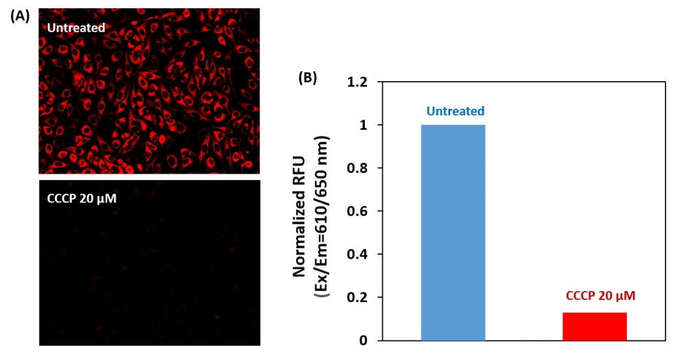
1.用测试化合物处理细胞一段时间,以诱导细胞凋亡,并建立平行对照实验。注意:我们用20 µM CCCP处理HeLa细胞15分钟,以改变线粒体膜电位。CCCP或FCCP可以与MitoTell Red同时添加。为了获得结果,可能需要为每个单独的细胞系滴定CCCP或FCCP。

2.将100 µL /孔/ 96孔板或25 µL /孔/ 384孔板的MitoTell Red工作溶液加入细胞板。

3.在避光的条件下,将板在37ºC下孵育15-30分钟。注意:适当的孵育时间取决于所用的单个细胞类型和细胞浓度。优化每个实验的孵育时间。

4.将50 µL /孔/ 96孔板或12.5 µL /孔/ 384孔板的测定缓冲液B(组分C)添加到细胞板中。注意:加载后请勿洗涤细胞。对于非贴壁细胞,建议在加入测定缓冲液B(组分C)后,以800 rpm离心细胞板2分钟,然后制动。

5.加入测定缓冲液B(组分C)后10到30分钟,用荧光酶标仪(Ext / Em = 610/650 nm(截止= 630 nm))检测荧光强度,或在荧光下用带Cy5滤镜的显微镜观察荧光信号。


参考文献

Safranine O as a fluorescent probe for mitochondrial membrane potential studied on the single particle level and in suspension
Authors: Perevoshchikova IV, Sorochkina AI, Zorov DB, Antonenko YN.
Journal: Biochemistry (Mosc) (2009): 663

Computer-assisted live cell analysis of mitochondrial membrane potential, morphology and calcium handling
Authors: Koopman WJ, Distelmaier F, Esseling JJ, Smeitink JA, Willems PH.
Journal: Methods (2008): 304

Determination of high mitochondrial membrane potential in spermatozoa loaded with the mitochondrial probe 5,5′,6,6′-tetrachloro-1,1′,3,3′-tetraethylbenzimidazolyl-carbocyanine iodide (JC-1) by using fluorescence-activated flow cytometry
Authors: Guthrie HD, Welch GR.
Journal: Methods Mol Biol (2008): 89

Effects of eprosartan on mitochondrial membrane potential and H2O2 levels in leucocytes in hypertension
Authors: Labios M, Martinez M, Gabriel F, Guiral V, Ruiz-Aja S, Beltran B, Munoz A.
Journal: J Hum Hypertens (2008): 493

Evaluation of sperm mitochondrial membrane potential by JC-1 fluorescent staining and flow cytometry
Authors: Xia XY, Wu YM, Hou BS, Yang B, Pan LJ, Shi YC, Jin BF, Shao Y, Cui YX, Huang YF.
Journal: Zhonghua Nan Ke Xue (2008): 135

How DASPMI reveals mitochondrial membrane potential: fluorescence decay kinetics and steady-state anisotropy in living cells
Authors: Ramadass R, Bereiter-Hahn J.
Journal: Biophys J (2008): 4068

Life cell quantification of mitochondrial membrane potential at the single organelle level
Authors: Distelmaier F, Koopman WJ, Testa ER, de Jong AS, Swarts HG, Mayatepek E, Smeitink JA, Willems PH.
Journal: Cytometry A (2008): 129

Mitochondrial membrane potential in axons increases with local nerve growth factor or semaphorin signaling
Authors: Verburg J, Hollenbeck PJ.
Journal: J Neurosci (2008): 8306

The mitochondrial membrane potential and Ca2+ oscillations in smooth muscle
Authors: Chalmers S, McCarron JG.
Journal: J Cell Sci (2008): 75

Cyclosporin A-induced oxidative stress is not the consequence of an increase in mitochondrial membrane potential
Authors: van der Toorn M, Kauffman HF, van der Deen M, Slebos DJ, Koeter GH, Gans RO, Bakker SJ.
Journal: Febs J (2007): 3003

Cell Meter 细胞内谷胱甘肽GSH检测试剂盒 适合流式细胞检测-AAT Bioquest荧光染料

上海金畔生物科技有限公司代理AAT Bioquest荧光染料全线产品,欢迎访问AAT Bioquest荧光染料官网了解更多信息。
Cell Meter 细胞内谷胱甘肽GSH检测试剂盒 适合流式细胞检测 价格 2823
产品规格

100 Tests

产品货号

Cell Meter 细胞内谷胱甘肽GSH检测试剂盒 适合流式细胞检测

产品参数
Ex (nm) 505 Em (nm) 524
分子量 溶剂
存储条件
产品概述

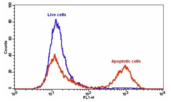
我们的Cell Meter 检测试剂盒是一套用于检测细胞生存力的工具。有多种参数可用于检测细胞活力。该特定试剂盒旨在通过测量耗尽的抗氧化剂还原型谷胱甘肽(GSH)来检测细胞凋亡。 GSH参与许多细胞过程,包括自由基,药物解毒,细胞信号传导和细胞增殖。细胞中GSH浓度的降低是许多细胞类型中对不同凋亡刺激物响应的细胞死亡进程的早期标志。我们的Cell Meter 细胞内GSH检测试剂盒为所有必需成分提供了优化的检测方法,用于检测GSH降低的细胞凋亡。该荧光测定法是基于使用我们专有的非荧光Thiolite Green染料检测细胞中GSH的结果,该染料与硫醇反应后会发出强烈的荧光。在正常细胞中,Thiolite Green主要在细胞质中积累,但在凋亡细胞中它会部分转移到线粒体中,而Thiolite Green的染色强度会降低。 Thiolite Green染色的细胞可以通过流式细胞仪观察。该试剂盒可与其他试剂配对,例如7-AAD(Cat#17501),碘化丙啶(Cat#17517),用于多参数研究细胞生存力和凋亡。该试剂盒经过优化,可通过流式细胞仪筛选凋亡抑制剂。金畔生物是AAT Bioquest的中国代理商,为您提供优质的Cell Meter 细胞内谷胱甘肽GSH检测试剂盒。

点击查看光谱


适用仪器


流式细胞仪  
Ex: 488 nm
Em: 530/30  nm 
通道: FITC 通道
实验方案

样品实验方案

简要概述

  1. 用5×10⁵至1×10⁶细胞/ mL的密度制备含测试化合物的细胞
  2. 将5 µL 200X Thiolite Green加入1 mL细胞溶液中
  3. 将细胞在37°C,5%CO2培养箱中孵育15至30分钟
  4. 沉淀细胞并将细胞重悬于1 mL生长培养基中
  5. 使用带有FL1通道的流式细胞仪分析细胞


溶液配制

储存溶液配制

1. Thiolite 绿色原液(200X):将500µL DMSO(组分C)加入小瓶Thiolite Green(组分A)中并充分混合,制成200X Thiolite Green储备液,避光。


实验步骤

1.对于每个样品,在1 mL温热培养基或自备缓冲液中以5×10⁵至1×10⁶细胞/ mL的密度制备细胞。注意:应单独评估每种细胞系,以确定诱导凋亡的细胞密度。

2.用测试化合物处理细胞所需的时间以诱导凋亡。

3.向处理过的细胞中加入5µL 200X Thiolite Green储备溶液。

4.将细胞在37°C,5%CO2培养箱中孵育15至30分钟。注意:对于贴壁细胞,用0.5 mM EDTA轻轻提起细胞以保持细胞完整,并在与Thiolite Green染料上样溶液孵育之前,用含血清的培养基清洗细胞一次。合适的孵育时间取决于所用的单个细胞类型和细胞浓度。优化每个实验的孵育时间。

5.可选:将细胞以1000 rpm离心4分钟,然后将细胞重悬于1 mL的测定缓冲液(组分B)或自备缓冲液中。

6.使用带有FL1通道的流式细胞仪(Ex / Em = 490/525 nm)检测荧光强度。


参考文献

The Combination of Astragalus membranaceus and Angelica sinensis Inhibits Lung Cancer and Cachexia through Its Immunomodulatory Function
Authors: Wu, Tsung-Han and Yeh, Kun-Yun and Wang, Chen-Hsu and Wang, Hang and Li, Tsung-Lin and Chan, Yi-Lin and Wu, Chang-Jer
Journal: Journal of oncology (2019)

ROS production and glutathione response in keratinocytes after application of β-carotene and VIS/NIR irradiation
Authors: Lohan, Silke B and Vitt, Kristina and Scholz, Patrik and Keck, Cornelia M and Meinke, Martina C
Journal: Chemico-biological interactions (2017)

Osmotic stress is accompanied by protein glycation in Arabidopsis thaliana
Authors: Paudel, Gagan and Bilova, Tatiana and Schmidt, Rico and Greifenhagen, Uta and Berger, Robert and Tarakhovskaya, Elena and Stöckhardt, Stefanie and Balcke, Gerd Ulrich and Humbeck, Klaus and Br and t, Wolfgang and others
Journal: Journal of Experimental Botany (2016): 6283–6295

Systemic Induction of NO-, Redox-, and cGMP Signaling in the Pumpkin Extrafascicular Phloem upon Local Leaf Wounding
Authors: Gaupels, Frank and Furch, Alex and ra CU and Zimmermann, Matthias R and Chen, Faxing and Kaever, Volkhard and Buhtz, Anja and Kehr, Julia and Sarioglu, Hakan and Kogel, Karl-Heinz and Durner, Jörg
Journal: Frontiers in plant science (2016)

Molecular mechanisms of hyperthermia-induced apoptosis enhanced by withaferin A
Authors: Cui, Zheng-Guo and Piao, Jin-Lan and Rehman, Mati UR and Ogawa, Ryohei and Li, Peng and Zhao, Qing-Li and Kondo, Takashi and Inadera, Hidekuni
Journal: European journal of pharmacology (2014): 99–107

Cancer & Metabolism
Authors: LaMonte, Gregory and Tang, Xiaohu and Chen, Julia Ling-Yu and Wu, Jianli and Ding, Chien-Kuang Cornelia and Keenan, Melissa M and Sangokoya, Carolyn and Kung, Hsiu-Ni and Ilkayeva, Olga and Boros, László G and others
Journal: (2013)

Cell Meter 通用Caspase活性荧光法检测试剂盒 绿色荧光 适合于流式细胞检测-AAT Bioquest荧光染料

上海金畔生物科技有限公司代理AAT Bioquest荧光染料全线产品,欢迎访问AAT Bioquest荧光染料官网了解更多信息。
Cell Meter 通用Caspase活性荧光法检测试剂盒 绿色荧光 适合于流式细胞检测 价格 2823
产品规格

100 Tests

产品货号

Cell Meter 通用Caspase活性荧光法检测试剂盒 绿色荧光 适合于流式细胞检测

产品参数
Ex (nm) 503 Em (nm) 525
分子量 溶剂
存储条件
产品概述

我们的Cell Meter 检测试剂盒是一套用于检测细胞生存力的工具。有多种参数可用于检测细胞活力。该特定试剂盒旨在通过测量活细胞中的通用胱天蛋白酶(胱天蛋白酶-1,-3,-4,-5,-6,-7,-8和-9)来检测细胞凋亡。胱天蛋白酶被广泛认为是细胞凋亡的可靠指标。大多数胱天蛋白酶具有对肽序列Val-Ala-Asp(VAD)的底物选择性。该试剂盒使用TF2-VAD-FMK作为大多数半胱天冬酶活性的荧光指示剂。TF2-VAD-FMK是细胞渗透性的,并且与凋亡细胞中活化的casepase-1,-3,-4,-5,-6,-7,-8和-9不可逆地结合。一旦与胱天蛋白酶结合,绿色荧光试剂将保留在细胞内。结合后阻止了胱天蛋白酶的进一步催化,但不会阻止凋亡的进行。添加到培养基中后的15分钟内,该试剂将开始与活性caspase酶反应。该试剂盒为所有必需成分提供了优化的测定方案。它用于定量凋亡细胞中大多数胱天蛋白酶活性,或筛选胱天蛋白酶抑制剂。绿色荧光标记允许通过流式细胞仪直接检测凋亡细胞胱天蛋白酶。金畔生物是AAT Bioquest的中国代理商,为您提供优质的Cell Meter 通用Caspase活性荧光法检测试剂盒

点击查看光谱


适用仪器


流式细胞仪  
Ex: 488 nm
Em: 530/30 nm
通道: FITC 通道
实验方案

样品实验方案

简要概述

  1. 用5×10⁵至1×10⁶细胞/ mL的密度制备含测试化合物的细胞
  2. 将0.5 µL细胞溶液中加入1 µL 500X TF2-VAD-FMK
  3. 将细胞在37°C,5%CO2培养箱中孵育1-4小时
  4. 沉淀细胞并将细胞重悬于0.5 mL检测缓冲液或生长培养基中
  5. 使用带有530/30 nm滤光片(FITC通道)的流式细胞仪分析细胞


实验步骤

1.对于每个样品,在0.5 mL温热培养基或自备缓冲液中以5×10⁵至1×10⁶cell/ mL的密度制备细胞。注意:应单独评估每种细胞系,以确定诱导凋亡的佳细胞密度。

2.用测试化合物处理细胞一段时间,以诱导细胞凋亡,并建立阳性和阴性对照。

3.向处理过的细胞中加入1µL 500X TF2-VAD-FMK(组分A)。

4.将细胞在37°C,5%CO2培养箱中孵育1-4小时。注意:对于贴壁细胞,用0.5 mM EDTA轻轻提起细胞以保持细胞完整,并在与TF2-VAD-FMK孵育之前用含血清的培养基洗涤细胞一次。合适的孵育时间取决于所用的单个细胞类型和细胞浓度。优化每个实验的孵育时间。

5.洗涤并旋转细胞两次。将细胞重悬于0.5 mL分析缓冲液(组分B)或生长培养基中。注意:TF2-VAD-FMK是荧光的;因此,重要的是洗净任何未结合的试剂以除去背景。

6.可选:可以用DNA染色剂标记细胞(例如碘化丙啶或死细胞的7-AAD)。

7.可选:修复细胞。

8.使用带有530/30 nm滤光片的流式细胞仪(FITC通道)检测荧光强度。


相关产品

Angiopoietins Modulate Survival, Migration, and the Components of the Ang-Tie2 Pathway of Chronic Lymphocytic Leukaemia (CLL) Cells In Vitro
Authors: Palma, Luis Mario Aguirre and Flamme, Hanna and Gerke, Iris and Kreuzer, Karl-Anton
Journal: Cancer Microenvironment (2016): 13–26

Hypoxia regulates TRAIL sensitivity of colorectal cancer cells through mitochondrial autophagy.
Authors: Knoll, Gertrud and Bittner, Sebastian and Kurz, Maria and Jantsch, Jonathan and Ehrenschwender, Martin
Journal: Oncotarget (2016)

Promotion of osteointegration under diabetic conditions by tantalum coating-based surface modification on 3-dimensional printed porous titanium implants
Authors: Wang, Lin and Hu, Xiaofan and Ma, Xiangyu and Ma, Zhensheng and Zhang, Yang and Lu, Yizhao and Li, Xiang and Lei, Wei and Feng, Yafei
Journal: Colloids and Surfaces B: Biointerfaces (2016): 440–452

Role of delta-like ligand-4 in chemoresistance against docetaxel in MCF-7 cells
Authors: Wang, Q and Shi, Y and Butler, HJ and Xue, J and Wang, G and Duan, P and Zheng, H
Journal: Human & Experimental Toxicology (2016): 0960327116650006

microRNA-186 inhibits cell proliferation and induces apoptosis in human esophageal squamous cell carcinoma by targeting SKP2
Authors: He, Wei and Feng, Jianfang and Zhang, Yan and Wang, Yuanyuan and Zang, Wenqiao and Zhao, Guoqiang
Journal: Laboratory Investigation (2016): 317–324

Glucose promotes cell proliferation, glucose uptake and invasion in endometrial cancer cells via AMPK/mTOR/S6 and MAPK signaling
Authors: Han, Jianjun and Zhang, Lu and Guo, Hui and Wysham, Weiya Z and Roque, Dario R and Willson, Adam K and Sheng, Xiugui and Zhou, Chunxiao and Bae-Jump, Victoria L
Journal: Gynecologic oncology (2015): 668–675

IL-7 abrogates the immunosuppressive function of human double-negative T cells by activating Akt/mTOR signaling
Authors: Allgäuer, Andrea and Schreiner, Elisabeth and Ferrazzi, Fulvia and Ekici, Arif B and Gerbitz, Armin and Mackensen, Andreas and Völkl, Simon
Journal: The Journal of Immunology (2015): 3139–3148

Insulin improves osteogenesis of titanium implants under diabetic conditions by inhibiting reactive oxygen species overproduction via the PI3K-Akt pathway
Authors: Wang, Lin and Zhao, Xiong and Wei, Bo-yuan and Liu, Yi and Ma, Xiang-yu and Wang, Jian and Cao, Peng-chong and Zhang, Yang and Yan, Ya-bo and Lei, Wei and others
Journal: Biochimie (2015): 85–93

LGL1 modulates proliferation, apoptosis, and migration of human fetal lung fibroblasts
Authors: Zhang, Hui and Sweezey, Neil B and Kaplan, Feige
Journal: American Journal of Physiology-Lung Cellular and Molecular Physiology (2015): L391–L402

t-BHQ Provides Protection against Lead Neurotoxicity via Nrf2/HO-1 Pathway
Authors: Ye, Fang and Li, Xiaoyi and Li, Lili and Yuan, Jing and Chen, Jun
Journal: Oxidative medicine and cellular longevity (2015)

Cell Meter 通用Caspase活性荧光法检测试剂盒 红色荧光 适合于流式细胞检测-AAT Bioquest荧光染料

上海金畔生物科技有限公司代理AAT Bioquest荧光染料全线产品,欢迎访问AAT Bioquest荧光染料官网了解更多信息。
Cell Meter 通用Caspase活性荧光法检测试剂盒 红色荧光 适合于流式细胞检测 价格 2823
产品规格

100 Tests

产品货号

Cell Meter 通用Caspase活性荧光法检测试剂盒 红色荧光 适合于流式细胞检测

产品参数
Ex (nm) 649 Em (nm) 664
分子量 溶剂
存储条件
产品概述

我们的Cell Meter 检测试剂盒是一套用于检测细胞生存力的工具。有多种参数可用于检测细胞活力。该特定试剂盒旨在通过测量活细胞中的通用胱天蛋白酶(胱天蛋白酶-1,-3,-4,-5,-6,-7,-8和-9)来检测细胞凋亡。胱天蛋白酶被广泛认为是细胞凋亡的可靠指标。大多数胱天蛋白酶具有对肽序列Val-Ala-Asp(VAD)的底物选择性。该试剂盒使用TF5-VAD-FMK作为大多数半胱天冬酶活性的荧光指示剂。 TF5-VAD-FMK是细胞可渗透的,并且与凋亡细胞中活化的casepase-1,-3,-4,-5,-6,-7,-8和-9不可逆地结合。一旦与胱天蛋白酶结合,红色荧光试剂将保留在细胞内。结合后阻止了胱天蛋白酶的进一步催化,但不会阻止凋亡的进行。添加到培养基中后的15分钟内,该试剂将开始与活性caspase酶反应。该试剂盒为所有必需成分提供了优化的测定方案。它用于定量凋亡细胞中大多数的胱天蛋白酶活性,或筛选胱天蛋白酶抑制剂。红色荧光标记允许通过流式细胞仪直接检测凋亡细胞中的胱天蛋白酶。金畔生物是AAT Bioquest的中国代理商,为您提供优质的Cell Meter 通用Caspase活性荧光法检测试剂盒。

点击查看光谱


适用仪器


流式细胞仪  
Ex: 640 nm
Em: 660/20 nm
通道: APC 通道
实验方案

样品实验方案

简要概述

  1. 用5×10⁵至1×10⁶细胞/ mL的密度制备含测试化合物的细胞
  2. 将0.5 µL细胞溶液中加入1 µL 500X TF5-VAD-FMK
  3. 将细胞在37°C,5%CO2培养箱中孵育1-4小时
  4. 沉淀细胞并将细胞重悬于0.5 mL检测缓冲液或生长培养基中
  5. 使用带有660/20 nm滤光片的流式细胞仪分析细胞(APC通道)


实验步骤

1.对于每个样品,在0.5mL温热培养基或自备缓冲液中以5×10⁵至1×10⁶cell/ mL的密度制备细胞。注意:应单独评估每种细胞系,以确定诱导凋亡的细胞密度。

2.用测试化合物处理细胞一段时间,以诱导细胞凋亡,并建立阳性和阴性对照。

3.向处理过的细胞中加入1µL 500X TF5-VAD-FMK(组分A)。

4.将细胞在37°C,5%CO2培养箱中孵育1-4小时。注意:对于贴壁细胞,用0.5 mM EDTA轻轻提起细胞以保持细胞完整,并在与TF5-VAD-FMK孵育之前用含血清的培养基洗涤细胞一次。合适的孵育时间取决于所用的单个细胞类型和细胞浓度。优化每个实验的孵育时间。

5.洗涤并旋转细胞两次。将细胞重悬于0.5 mL分析缓冲液(组分B)或生长培养基中。注意:TF5-VAD-FMK是荧光的;因此,重要的是洗净任何未结合的试剂以除去背景。

6.可选:用DNA染色剂标记细胞(例如绿色DCS标记死细胞,可以用530/30 nm滤光片(FITC通道)检测到。

7.可选:修复细胞。

8.使用带有660/20 nm滤光片(APC通道)的流式细胞仪检测荧光强度。


参考文献

Angiopoietins Modulate Survival, Migration, and the Components of the Ang-Tie2 Pathway of Chronic Lymphocytic Leukaemia (CLL) Cells In Vitro
Authors: Palma, Luis Mario Aguirre and Flamme, Hanna and Gerke, Iris and Kreuzer, Karl-Anton
Journal: Cancer Microenvironment (2016): 13–26

Hypoxia regulates TRAIL sensitivity of colorectal cancer cells through mitochondrial autophagy.
Authors: Knoll, Gertrud and Bittner, Sebastian and Kurz, Maria and Jantsch, Jonathan and Ehrenschwender, Martin
Journal: Oncotarget (2016)

Promotion of osteointegration under diabetic conditions by tantalum coating-based surface modification on 3-dimensional printed porous titanium implants
Authors: Wang, Lin and Hu, Xiaofan and Ma, Xiangyu and Ma, Zhensheng and Zhang, Yang and Lu, Yizhao and Li, Xiang and Lei, Wei and Feng, Yafei
Journal: Colloids and Surfaces B: Biointerfaces (2016): 440–452

Role of delta-like ligand-4 in chemoresistance against docetaxel in MCF-7 cells
Authors: Wang, Q and Shi, Y and Butler, HJ and Xue, J and Wang, G and Duan, P and Zheng, H
Journal: Human & Experimental Toxicology (2016): 0960327116650006

microRNA-186 inhibits cell proliferation and induces apoptosis in human esophageal squamous cell carcinoma by targeting SKP2
Authors: He, Wei and Feng, Jianfang and Zhang, Yan and Wang, Yuanyuan and Zang, Wenqiao and Zhao, Guoqiang
Journal: Laboratory Investigation (2016): 317–324

Glucose promotes cell proliferation, glucose uptake and invasion in endometrial cancer cells via AMPK/mTOR/S6 and MAPK signaling
Authors: Han, Jianjun and Zhang, Lu and Guo, Hui and Wysham, Weiya Z and Roque, Dario R and Willson, Adam K and Sheng, Xiugui and Zhou, Chunxiao and Bae-Jump, Victoria L
Journal: Gynecologic oncology (2015): 668–675

IL-7 abrogates the immunosuppressive function of human double-negative T cells by activating Akt/mTOR signaling
Authors: Allgäuer, Andrea and Schreiner, Elisabeth and Ferrazzi, Fulvia and Ekici, Arif B and Gerbitz, Armin and Mackensen, Andreas and Völkl, Simon
Journal: The Journal of Immunology (2015): 3139–3148

Insulin improves osteogenesis of titanium implants under diabetic conditions by inhibiting reactive oxygen species overproduction via the PI3K-Akt pathway
Authors: Wang, Lin and Zhao, Xiong and Wei, Bo-yuan and Liu, Yi and Ma, Xiang-yu and Wang, Jian and Cao, Peng-chong and Zhang, Yang and Yan, Ya-bo and Lei, Wei and others
Journal: Biochimie (2015): 85–93

LGL1 modulates proliferation, apoptosis, and migration of human fetal lung fibroblasts
Authors: Zhang, Hui and Sweezey, Neil B and Kaplan, Feige
Journal: American Journal of Physiology-Lung Cellular and Molecular Physiology (2015): L391–L402

t-BHQ Provides Protection against Lead Neurotoxicity via Nrf2/HO-1 Pathway
Authors: Ye, Fang and Li, Xiaoyi and Li, Lili and Yuan, Jing and Chen, Jun
Journal: Oxidative medicine and cellular longevity (2015)

Cell Meter Caspase 3/7活性细胞凋亡检测试剂盒 绿色荧光 适合于流式细胞检测-AAT Bioquest荧光染料

上海金畔生物科技有限公司代理AAT Bioquest荧光染料全线产品,欢迎访问AAT Bioquest荧光染料官网了解更多信息。
Cell Meter Caspase 3/7活性细胞凋亡检测试剂盒 绿色荧光 适合于流式细胞检测 价格 2823
产品规格

100 Tests

产品货号

Cell Meter Caspase 3/7活性细胞凋亡检测试剂盒 绿色荧光 适合于流式细胞检测

产品参数
Ex (nm) 503 Em (nm) 525
分子量 溶剂
存储条件
产品概述

Cell Meter Caspase 3/7活性细胞凋亡检测试剂盒是美国AAT Bioquest生产的用于细胞凋亡检测的试剂盒,Cell Meter检测试剂盒是一类用于检测细胞功能的系列工具,包括细胞活性、细胞毒性、细胞凋亡、细胞膜电位以及细胞周期等方面的指标。每种检测方案均能提供不同荧光颜色的检测方案。这些检测方案为从多角度研究细胞功能活动提供了一种十分有效的方法。Cell Meter Caspase 3/7活性细胞凋亡检测试剂盒是通过测量Caspase 3/7的活性而用于细胞凋亡检测的。因为Caspase-3 (CPP32/apopain)在细胞凋亡的起始过程中发挥着重要作用,因此其作为一个值得信赖的细胞凋亡指示剂广泛地被大家所接受。Caspase 3具有特异性的底物多肽识别序列,即天冬氨酸-谷氨酸-缬氨酸-天冬氨酸(DEVD)。本试剂盒用TF2-DEVD-FMK作为荧光指示剂,用于Caspase-3/7活性的检测。TF2-DEVD-FMK具有细胞渗透性、无细胞毒性,在凋亡细胞中能与活化的casepase-3/7进行不可逆的结合。一旦与Caspases结合,这种绿色荧光染料会被保留在细胞中。这种结合可以阻止Caspases进一步的催化作用,但是不会使细胞凋亡的进程停止。这种试剂将在添加介质之后的15分钟内与有活性的Caspase酶进行反应。本试剂盒提供所有必需组分和检测方案。它用来对凋亡细胞中绝大多数活化的Caspases的活性进行定量检测,或者用来筛选Caspases抑制剂。这种绿色荧光可以通过流式细胞仪对的Caspases进行直接检测。金畔生物是AAT Bioquest的中国代理商,为您提供优质的Cell Meter Caspase 3/7活性细胞凋亡检测试剂盒。 

点击查看光谱


适用仪器


流式细胞仪  
Ex: 488 nm
Em: 530/30 nm 
通道: FITC 通道
实验方案

样品实验方案

简要概述

1.用测试化合物制备细胞,密度为5×10⁵至1×10⁶个细胞/ mL
2.将1μL500XTF2-DEVD-FMK加入0.5mL细胞溶液中
3.在37°C,5%CO2培养箱中孵育1-4小时
4.将细胞沉淀并将细胞重悬于0.5mL测定缓冲液或生长培养基中
5.使用具有FL1通道的流式细胞仪分析细胞


操作步骤

1.对于每个样品,准备细胞在0.5 mL温热培养基或您选择的缓冲液中,密度为5×10⁵至1×10⁶个细胞/ mL。注意:应对每个细胞系进行单独评估,以确定诱导细胞凋亡的佳细胞密度。

2.用测试化合物处理细胞一段所需的时间以诱导细胞凋亡,并产生阳性和阴性对照。

3.将1μL500XTF2-DEVD-FMK(组分A)加入处理过的细胞中。

4.将细胞在37°C,5%CO2培养箱中孵育1-4小时。注意:对于贴壁细胞,用0.5 mM EDTA轻轻提起细胞以保持细胞完整,并在用TF2-DEVD-FMK孵育之前用含血清的培养基洗涤细胞一次。适当的孵育时间取决于所用的单个细胞类型和细胞浓度。优化每个实验的孵育时间。

5.将细胞洗涤并旋转两次。将细胞重悬于0.5mL测定缓冲液或生长培养基中。注意:TF2-DEVD-FMK是荧光的;因此,任何未结合的试剂以去除背景是很重要的。

6.用DNA染色标记细胞(如碘化丙锭或7-AAD用于死细胞)。

7.使用具有FL1通道的流式细胞仪(Ex / Em = 490 / 525nm)监测荧光强度。


数据分析

Cell Meter Caspase 3/7活性细胞凋亡检测试剂盒 绿色荧光 适合于流式细胞检测

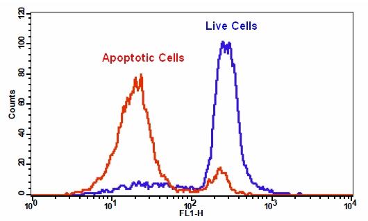
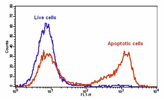
图1.使用Jurkat细胞中的Cell Meter Caspase 3/7活性细胞凋亡检测试剂盒检测caspase 3/7活性。 加入喜树碱诱导TF2-DEVD-FMK荧光强度。 将Jurkat细胞在37℃,5%CO2培养箱中不用(蓝色)或用20μM喜树碱(红色)处理4-5小时,然后用TF2-DEVD-FMK染色1小时。


参考文献

Angiopoietins Modulate Survival, Migration, and the Components of the Ang-Tie2 Pathway of Chronic Lymphocytic Leukaemia (CLL) Cells In Vitro
Authors: Luis Mario Aguirre Palma, Hanna Flamme, Iris Gerke, Karl-Anton Kreuzer
Journal: Cancer Microenvironment (2016): 13–26

Hypoxia regulates TRAIL sensitivity of colorectal cancer cells through mitochondrial autophagy.
Authors: Gertrud Knoll, Sebastian Bittner, Maria Kurz, Jonathan Jantsch, Martin Ehrenschwender
Journal: Oncotarget (2016)

microRNA-186 inhibits cell proliferation and induces apoptosis in human esophageal squamous cell carcinoma by targeting SKP2
Authors: Wei He, Jianfang Feng, Yan Zhang, Yuanyuan Wang, Wenqiao Zang, Guoqiang Zhao
Journal: Laboratory Investigation (2016): 317–324

Promotion of osteointegration under diabetic conditions by tantalum coating-based surface modification on 3-dimensional printed porous titanium implants
Authors: Lin Wang, Xiaofan Hu, Xiangyu Ma, Zhensheng Ma, Yang Zhang, Yizhao Lu, Xiang Li, Wei Lei, Yafei Feng
Journal: Colloids and Surfaces B: Biointerfaces (2016): 440–452

Role of delta-like ligand-4 in chemoresistance against docetaxel in MCF-7 cells
Authors: Q Wang, Y Shi, HJ Butler, J Xue, G Wang, P Duan, H Zheng
Journal: Human & Experimental Toxicology (2016): 0960327116650006

Glucose promotes cell proliferation, glucose uptake and invasion in endometrial cancer cells via AMPK/mTOR/S6 and MAPK signaling
Authors: Jianjun Han, Lu Zhang, Hui Guo, Weiya Z Wysham, Dario R Roque, Adam K Willson, Xiugui Sheng, Chunxiao Zhou, Victoria L Bae-Jump
Journal: Gynecologic oncology (2015): 668–675

IL-7 abrogates the immunosuppressive function of human double-negative T cells by activating Akt/mTOR signaling
Authors: Andrea Allgäuer, Elisabeth Schreiner, Fulvia Ferrazzi, Arif B Ekici, Armin Gerbitz, Andreas Mackensen, Simon Völkl
Journal: The Journal of Immunology (2015): 3139–3148

Insulin improves osteogenesis of titanium implants under diabetic conditions by inhibiting reactive oxygen species overproduction via the PI3K-Akt pathway
Authors: Lin Wang, Xiong Zhao, Bo-yuan Wei, Yi Liu, Xiang-yu Ma, Jian Wang, Peng-chong Cao, Yang Zhang, Ya-bo Yan, Wei Lei
Journal: Biochimie (2015): 85–93

LGL1 modulates proliferation, apoptosis, and migration of human fetal lung fibroblasts
Authors: Hui Zhang, Neil B Sweezey, Feige Kaplan
Journal: American Journal of Physiology-Lung Cellular and Molecular Physiology (2015): L391–L402

t-BHQ Provides Protection against Lead Neurotoxicity via Nrf2/HO-1 Pathway
Authors: Fang Ye, Xiaoyi Li, Lili Li, Jing Yuan, Jun Chen
Journal: Oxidative medicine and cellular longevity (2015)


相关产品

产品名称 货号
Cell Meter Caspase 3/7活性细胞凋亡检测试剂盒 蓝色荧光 Cat#22795
Cell Meter Caspase 3/7活性细胞凋亡检测试剂盒 绿色荧光 Cat#22796
Cell Meter Caspase 3/7活性细胞凋亡检测试剂盒 红色荧光 Cat#22797

Cell Meter Annexin V凋亡检测试剂盒 绿色荧光 适合流式细胞检测-AAT Bioquest荧光染料

上海金畔生物科技有限公司代理AAT Bioquest荧光染料全线产品,欢迎访问AAT Bioquest荧光染料官网了解更多信息。
Cell Meter Annexin V凋亡检测试剂盒 绿色荧光 适合流式细胞检测 价格 2823
产品规格

100 Tests

产品货号

Cell Meter Annexin V凋亡检测试剂盒 绿色荧光 适合流式细胞检测

产品参数
Ex (nm) 491 Em (nm) 516
分子量 溶剂
存储条件
产品概述

Cell Meter Annexin V凋亡检测试剂盒是美国AAT Bioquest生产的细胞凋亡检测试剂盒,我们的Cell Meter 检测试剂盒是一套用于检测细胞生存力的工具。有多种参数可用于检测细胞活力。该特定试剂盒旨在通过测量磷脂酰丝氨酸(PS)的转运来监测细胞凋亡。在凋亡中,PS转移到质膜的外部小叶。磷脂酰丝氨酸在细胞表面的出现是细胞凋亡初始/中间阶段的普遍指标,可以在观察到形态变化之前进行检测。该试剂盒使用与PS特异性结合的荧光膜联蛋白V。已经证明膜联蛋白V缀合物选择性结合PS,使用带有FITC通道的流式细胞仪(绿色荧光)优化了此特定的检测试剂盒,以检测细胞凋亡。金畔生物是AAT Bioquest的中国代理商,为您提供优质的Cell Meter Annexin V凋亡检测试剂盒。

点击查看光谱 


适用仪器


流式细胞仪  
Ex: 488 nm
Em: 530/30 nm 
通道: FITC 通道
实验方案

样品实验方案

简要概述

用测试化合物(200μL/样品)制备细胞
添加Annexin V-iFluor 488原液
在室温下孵育30-60分钟
使用FL1通道的流式细胞仪分析细胞(Ex / Em = 490/525 nm)


操作步骤

1.用测试化合物处理细胞一段时间(用喜树碱处理的Jurkat细胞4-6小时)以诱导细胞凋亡。注意:膜粘附细胞的膜联蛋白V流式细胞术分析未经常检测,因为在细胞分离或收获期间可能发生特定的膜损伤。然而,Casiola-Rosen等人先前报道了利用膜联蛋白V对贴壁细胞类型进行流式细胞术的方法。和van Engelend等人。

2.离心细胞,得到1-5×10⁵个细胞/管。

3.将细胞重悬于200μL测定缓冲液(组分B)中。

4.将2μL膜联蛋白V-iFluor 488(组分A)加入细胞中。

5.可选:加入2μL100X碘化丙啶(成分C)用于坏死细胞。

6.在室温下孵育30至60分钟,避光。

7.可选:在使用流式细胞仪分析细胞之前,添加200至300μL的测定缓冲液(组分B)以增加体积。

8.使用具有FL1通道的流式细胞仪(Ex / Em = 490 / 525nm)监测膜联蛋白V-iFluor 488的荧光强度。当碘化丙啶加入细胞时,使用FL2通道测量细胞活力。


数据分析

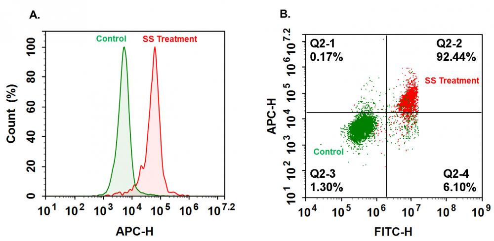
        在活的非凋亡细胞中,膜联蛋白V-iFluor 488检测非诱导细胞中的先天性细胞凋亡,其通常为所有细胞的2-6%。 在凋亡细胞中,膜联蛋白V-iFluor 488与磷脂酰丝氨酸结合,磷脂酰丝氨酸位于细胞膜的外部小叶上,因此导致染色强度增加。

Cell Meter Annexin V凋亡检测试剂盒 绿色荧光 适合流式细胞检测

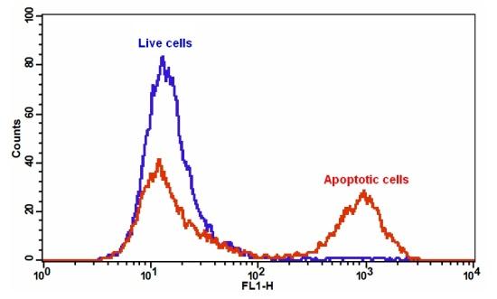
图1.检测膜联蛋白V-iFluor 488和磷脂酰丝氨酸在Jurkat细胞中的结合活性。 Jurkat细胞在37℃,5%CO2培养箱中不用(蓝色)或用20μM喜树碱(红色)处理4-5小时,然后用Annexin V-iFluor 488染色30分钟。 使用FL1通道用FACSCalibur(Becton Dickinson)流式细胞仪测量膜联蛋白V-iFluor 488的荧光强度。


参考文献

Combined Treatments of Magnetic Intra-Lysosomal Hyperthermia with Doxorubicin Promotes Synergistic Anti-Tumoral Activity.
Authors: D Hajj Diab El, P Clerc, N Serhan, D Fourmy, V Gigoux
Journal: Nanomaterials (Basel, Switzerland) (2018)

In situ polymerizable hydrogel incorporated with specific pathogen-free porcine platelet-rich plasma for the reconstruction of the corneal endothelium
Authors: Yung-Kai Lin, Ruchi Sharma, Hsu Ma, Wen-Shyan Chen, Chao-Ling Yao
Journal: Journal of the Taiwan Institute of Chemical Engineers (2017)

Novel regulations of MEF2-A, MEF2-D, and CACNA1S in the functional incompetence of adipose-derived mesenchymal stem cells by induced indoxyl sulfate in chronic kidney disease
Authors: Duyen Thi Do, Nam Nhut Phan, Chih-Yang Wang, Zhengda Sun, Yen-Chang Lin
Journal: Cytotechnology (2016): 2589–2604

Shear stress-induced alteration of epithelial organization in human renal tubular cells
Authors: Damien Maggiorani, Romain Dissard, Marcy Belloy, Jean-Sébastien Saulnier-Blache, Audrey Casemayou, Laure Ducasse, Sandra Grès, Julie Bellière, Cécile Caubet, Jean-Loup Bascands
Journal: PloS one (2015): e0131416

Targeting a G-protein-coupled receptor overexpressed in endocrine tumors by magnetic nanoparticles to induce cell death
Authors: Claire Sanchez, Darine El Hajj Diab, Vincent Connord, Pascal Clerc, Etienne Meunier, Bernard Pipy, Bruno Payré, Reasmey P Tan, Michel Gougeon, Julian Carrey
Journal: Acs Nano (2014): 1350–1363


相关产品

产品名称 货号
Cell Meter Annexin V凋亡检测试剂盒 红色荧光 适合流式细胞检测 Cat#22826
Cell Meter Annexin V凋亡检测试剂盒 深红色荧光 适合流式细胞检测 Cat#22827
Cell Meter Annexin V凋亡检测试剂盒 橙色荧光 适合流式细胞检测 Cat#22825

Cell Meter Annexin V凋亡检测试剂盒 橙色荧光 适合流式细胞检测-AAT Bioquest荧光染料

上海金畔生物科技有限公司代理AAT Bioquest荧光染料全线产品,欢迎访问AAT Bioquest荧光染料官网了解更多信息。
Cell Meter Annexin V凋亡检测试剂盒 橙色荧光 适合流式细胞检测 价格 2823
产品规格

100 Tests

产品货号

Cell Meter Annexin V凋亡检测试剂盒 橙色荧光 适合流式细胞检测

产品参数
Ex (nm) 557 Em (nm) 570
分子量 溶剂
存储条件
产品概述

我们的Cell Meter 检测试剂盒是一套用于检测细胞生存力的工具,有多种参数可用于检测细胞活力。 该特定试剂盒旨在通过测量磷脂酰丝氨酸(PS)的转运来检测细胞凋亡。 在细胞凋亡中,PS转移到质膜的外部小叶。 磷脂酰丝氨酸在细胞表面的出现是细胞凋亡初始/中间阶段的普遍指标,可以在观察形态变化之前进行检测。该试剂盒使用与PS特异性结合的荧光膜联蛋白V。 已经证明膜联蛋白V缀合物选择性结合PS。 使用带有575/26 nm发射滤光片组(PE通道)的流式细胞仪,优化了该特定的测定试剂盒以监测细胞凋亡。金畔生物是AAT Bioquest的中国代理商,为您提供优质的Cell Meter Annexin V凋亡检测试剂盒。

点击查看光谱


适用仪器


流式细胞仪  
Ex: 488 nm or 532 nm
Em: 575/26 nm 
通道: PE 通道
实验方案

样品实验方案

简要概述

  1. 用测试化合物制备细胞(200 µL /样品)
  2. 添加膜联蛋白V-iFluor 555测定溶液
  3. 在室温下孵育30至60分钟
  4. 使用带有575/26 nm滤光片(PE通道)的流式细胞仪或带有Cy3滤光片组的荧光显微镜分析细胞


实验步骤

1.用膜联蛋白V-iFluor 555准备和孵育细胞:

1.1用测试化合物处理细胞一段时间(对于用星形孢菌素处理过的Jurkat细胞,需要4-6小时)以诱导细胞凋亡。

1.2离心细胞以获得1-5×10⁵细胞/管。

1.3将细胞重悬于200 µL分析缓冲液(组分B)中。

1.4向细胞中加入2 µL Annexin V-iFluor 555(组分A)。

1.5避光保存,于室温下孵育30至60分钟。

1.6在使用流式细胞仪或荧光显微镜分析细胞之前,添加300 µL分析缓冲液(组分B)以增加体积。

1.7使用带有575/26 nm滤光片(PE通道)的流式细胞仪或带有Cy3滤光片组的荧光显微镜检测荧光强度。

2.通过使用流式细胞仪进行分析:

2.1使用带有575/26 nm滤光片的流式细胞仪(PE通道)定量Annexin V- iFluor 555结合物。

3.通过使用荧光显微镜进行分析:

3.1孵育后用移液管吸移细胞悬液,用测定缓冲液冲洗1-2次,然后用测定缓冲液重悬细胞。

3.2将细胞添加到载玻片盖玻片的载玻片上。注意:对于贴壁细胞,建议直接在盖玻片上生长细胞。与Annexin V-iFluor 555孵育后,用测定缓冲液冲洗1-2次,然后将测定缓冲液加到盖玻片上。将玻片上的盖玻片倒置并可视化细胞。与膜联蛋白V-iFluor 555孵育后,还可以将细胞固定在2%甲醛中,并在显微镜下观察。

3.3在荧光显微镜下使用Cy3滤光片组,用Annexin V-iFluor 555分析凋亡细胞。将Nuclear Red DCS1(#17552)添加到细胞后,使用Cy5通道测量细胞活力。质膜上的橙色染色表明膜联蛋白V-iFluor 555与细胞表面的PS结合。


参考文献

In situ polymerizable hydrogel incorporated with specific pathogen-free porcine platelet-rich plasma for the reconstruction of the corneal endothelium
Authors: Lin, Yung-Kai and Sharma, Ruchi and Ma, Hsu and Chen, Wen-Shyan and Yao, Chao-Ling
Journal: Journal of the Taiwan Institute of Chemical Engineers (2017)

Novel regulations of MEF2-A, MEF2-D, and CACNA1S in the functional incompetence of adipose-derived mesenchymal stem cells by induced indoxyl sulfate in chronic kidney disease
Authors: Do, Duyen Thi and Phan, Nam Nhut and Wang, Chih-Yang and Sun, Zhengda and Lin, Yen-Chang
Journal: Cytotechnology (2016): 2589–2604

Shear stress-induced alteration of epithelial organization in human renal tubular cells
Authors: Maggiorani, Damien and Dissard, Romain and Belloy, Marcy and Saulnier-Blache, Jean-Sébastien and Casemayou, Audrey and Ducasse, Laure and Grès, S and ra and Bellière, Julie and Caubet, Cécile and Basc and s, Jean-Loup and others
Journal: PloS one (2015): e0131416

Targeting a G-protein-coupled receptor overexpressed in endocrine tumors by magnetic nanoparticles to induce cell death
Authors: Sanchez, Claire and El Hajj Diab, Darine and Connord, Vincent and Clerc, Pascal and Meunier, Etienne and Pipy, Bernard and Payré, Bruno and Tan, Reasmey P and Gougeon, Michel and Carrey, Julian and others
Journal: Acs Nano (2014): 1350–1363

Glycogen synthase kinase-3 and Omi/HtrA2 induce annexin A2 cleavage followed by cell cycle inhibition and apoptosis
Authors: Wang CY, Lin YS, Su WC, Chen CL, Lin CF.
Journal: Mol Biol Cell (2009): 4153

Gold fluorescent annexin A5 as a novel apoptosis detection tool
Authors: Kurschus FC, Pal PP, Baumler P, Jenne DE, Wiltschi B, Budisa N.
Journal: Cytometry A (2009): 626

Detection of apoptosis induced by new type gosling viral enteritis virus in vitro through fluorescein annexin V-FITC/PI double labeling
Authors: Chen S, Cheng AC, Wang MS, Peng X.
Journal: World J Gastroenterol (2008): 2174

Evaluation of annexin V and Calcein-AM as markers of mononuclear cell apoptosis during human immunodeficiency virus infection
Authors: Palma PF, Baggio GL, Spada C, Silva RD, Ferreira SI, Treitinger A.
Journal: Braz J Infect Dis (2008): 108

Measurement of annexin V uptake and lactadherin labeling for the quantification of apoptosis in adherent Tca8113 and ACC-2 cells
Authors: Hu T, Shi J, Jiao X, Zhou J, Yin X.
Journal: Braz J Med Biol Res (2008): 750

Rhodamine B isothiocyanate doped silica-coated fluorescent nanoparticles (RBITC-DSFNPs)-based bioprobes conjugated to Annexin V for apoptosis detection and imaging
Authors: Shi H, He X, Wang K, Yuan Y, Deng K, Chen J, Tan W.
Journal: Nanomedicine (2007): 266

Cell Meter Annexin V凋亡检测试剂盒 红色荧光 适合流式细胞检测-AAT Bioquest荧光染料

上海金畔生物科技有限公司代理AAT Bioquest荧光染料全线产品,欢迎访问AAT Bioquest荧光染料官网了解更多信息。
Cell Meter Annexin V凋亡检测试剂盒 红色荧光 适合流式细胞检测 价格 2823
产品规格

100 Tests

产品货号

Cell Meter Annexin V凋亡检测试剂盒 红色荧光 适合流式细胞检测

产品参数
Ex (nm) 587 Em (nm) 603
分子量 溶剂
存储条件
产品概述

Cell Meter Annexin V凋亡检测试剂盒是美国AAT Bioquest生产的用于检测细胞凋亡的试剂盒,我们的Cell Meter 检测试剂盒是一套用于检测细胞生存力的工具。有多种参数可用于检测细胞活力。该特定试剂盒旨在通过测量磷脂酰丝氨酸(PS)的转运来监测细胞凋亡。在凋亡中,PS转移到质膜的外部小叶。磷脂酰丝氨酸在细胞表面的出现是细胞凋亡初始/中间阶段的普遍指标,可以在观察到形态变化之前进行检测。该试剂盒使用与PS特异性结合的荧光膜联蛋白V。已经证明膜联蛋白V缀合物选择性结合PS,使用带有PE-Texas Red通道的流式细胞仪(红色荧光)优化了此特定的检测试剂盒,以检测细胞凋亡。金畔生物是AAT Bioquest的中国代理商,为您提供优质的Cell Meter Annexin V凋亡检测试剂盒。 

点击查看光谱


适用仪器


流式细胞仪  
Ex: 561 nm
Em: 610/20  nm 
通道: PE-Texas Red 通道
实验方案

样品实验方案

简要概述

1.用测试化合物(200μL/样品)制备细胞
2.添加Annexin V-iFluor 594检测溶液
3.在室温下孵育30-60分钟
4.使用具有Ex / Em = 590 / 620nm滤光片组的流式细胞仪或具有TRITC或Texas Red通道的荧光显微镜分析细胞


操作步骤

使用Annexin V-iFluor 594制备和孵育细胞:

1.用测试化合物处理细胞一段所需的时间(用星形孢菌素处理的Jurkat细胞4-6小时)以诱导细胞凋亡。

2.离心细胞,得到1-5×10⁵个细胞/管。

3.将细胞重悬于200μL测定缓冲液(组分B)中。

4.将2μL膜联蛋白V-iFluor 594(组分A)加入细胞中。

5.在室温下孵育30至60分钟,避光。

6.在使用流式细胞仪或荧光显微镜分析细胞之前,添加300μL分析缓冲液(组分B)以增加体积。

7.使用具有Ex / Em = 590 / 620nm滤光器组的流式细胞仪或具有TRITC或Texas Red通道的荧光显微镜监测荧光强度。


使用流式细胞仪分析:

使用流式细胞仪在Ex / Em = 590 / 620nm处定量膜联蛋白V-iFluor 594结合。 注意:粘附细胞上的膜联蛋白V结合流式细胞术分析未经常检测,因为在细胞分离或收获期间可能发生特定的膜损伤。 然而,Casciola-Rosen等人先前报道了利用膜联蛋白V对贴壁细胞类型进行流式细胞术的方法。 


使用荧光显微镜分析:

1.孵育后移取细胞悬液,用测定缓冲液冲洗1-2次,然后用测定缓冲液重悬细胞。

2.将细胞添加到盖有玻璃盖玻片的载玻片上。 注意:对于粘附细胞,建议直接在盖玻片上生长细胞。 与Annexin V-iFluor 594孵育后,用测定缓冲液冲洗1-2次,并将测定缓冲液加回到盖玻片中。 在玻璃载玻片上翻转盖玻片并可视化细胞。 在与膜联蛋白V-iFluor 594孵育后,细胞也可以固定在2%甲醛中,并在显微镜下观察。

3.使用TRITC或Texas Red通道在荧光显微镜下用膜联蛋白V-iFluor 594分析凋亡细胞。 质膜上的橙色染色表明膜联蛋白V-iFluor 594与细胞表面上的PS结合。


数据分析

        在活的非凋亡细胞中,膜联蛋白V-iFluor 594检测非诱导细胞中的先天性细胞凋亡,其通常为所有细胞的2-6%。 在凋亡细胞中,膜联蛋白V-iFluor 594与磷脂酰丝氨酸结合,磷脂酰丝氨酸位于细胞膜的外部小叶上,导致染色强度增加。

Cell Meter Annexin V凋亡检测试剂盒 红色荧光 适合流式细胞检测

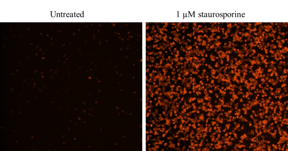
图1.用细胞仪膜联蛋白V结合细胞凋亡检测试剂盒染色的Costar黑壁/透明底96孔板中Jurkat细胞的图像*红色荧光*。 (左):未处理的对照细胞。 (右):用1μM星形孢菌素处理细胞5小时。


参考文献

In situ polymerizable hydrogel incorporated with specific pathogen-free porcine platelet-rich plasma for the reconstruction of the corneal endothelium
Authors: Yung-Kai Lin, Ruchi Sharma, Hsu Ma, Wen-Shyan Chen, Chao-Ling Yao
Journal: Journal of the Taiwan Institute of Chemical Engineers (2017)

Novel regulations of MEF2-A, MEF2-D, and CACNA1S in the functional incompetence of adipose-derived mesenchymal stem cells by induced indoxyl sulfate in chronic kidney disease
Authors: Duyen Thi Do, Nam Nhut Phan, Chih-Yang Wang, Zhengda Sun, Yen-Chang Lin
Journal: Cytotechnology (2016): 2589–2604

Shear stress-induced alteration of epithelial organization in human renal tubular cells
Authors: Damien Maggiorani, Romain Dissard, Marcy Belloy, Jean-Sébastien Saulnier-Blache, Audrey Casemayou, Laure Ducasse, Sandra Grès, Julie Bellière, Cécile Caubet, Jean-Loup Bascands
Journal: PloS one (2015): e0131416

Targeting a G-protein-coupled receptor overexpressed in endocrine tumors by magnetic nanoparticles to induce cell death
Authors: Claire Sanchez, Darine El Hajj Diab, Vincent Connord, Pascal Clerc, Etienne Meunier, Bernard Pipy, Bruno Payré, Reasmey P Tan, Michel Gougeon, Julian Carrey
Journal: Acs Nano (2014): 1350–1363


相关产品

产品名称 货号
Cell Meter Annexin V凋亡检测试剂盒 绿色荧光 适合流式细胞检测 Cat#22824
Cell Meter Annexin V凋亡检测试剂盒 深红色荧光 适合流式细胞检测 Cat#22827
Cell Meter Annexin V凋亡检测试剂盒 橙色荧光 适合流式细胞检测 Cat#22825

Cell Meter Annexin V凋亡检测试剂盒 深红色荧光 适合流式细胞检测-AAT Bioquest荧光染料

上海金畔生物科技有限公司代理AAT Bioquest荧光染料全线产品,欢迎访问AAT Bioquest荧光染料官网了解更多信息。
Cell Meter Annexin V凋亡检测试剂盒 深红色荧光 适合流式细胞检测 价格 2823
产品规格

100 Tests

产品货号

Cell Meter Annexin V凋亡检测试剂盒 深红色荧光 适合流式细胞检测

产品参数
Ex (nm) 656 Em (nm) 670
分子量 溶剂
存储条件
产品概述

我们的Cell Meter 检测试剂盒是一套用于检测细胞生存力的工具,有多种参数可用于监测细胞活力。 该特定试剂盒旨在通过测量磷脂酰丝氨酸(PS)的转运来检测细胞凋亡。 在细胞凋亡中,PS转移到质膜的外部小叶。 磷脂酰丝氨酸在细胞表面的出现是细胞凋亡初始/中间阶段的普遍指标,可以在观察形态变化之前进行检测。 该试剂盒使用与PS特异性结合的荧光膜联蛋白V。 已经证明膜联蛋白V缀合物选择性结合PS。 使用带有Cy5滤波片的流式细胞仪优化了该特定的检测试剂盒,以检测细胞凋亡。金畔生物是AAT Bioquest的中国代理商,为您提供优质的Cell Meter Annexin V凋亡检测试剂盒。

点击查看光谱


适用仪器


流式细胞仪  
Ex: 640 nm
Em: 660/20 nm
通道: APC 通道
实验方案

样品实验方案

简要概述

  1. 用测试化合物制备细胞(200 µL /样品)
  2. 添加膜联蛋白V-iFluor 647测定溶液
  3. 在室温下孵育30-60分钟
  4. 使用带有660/20 nm滤光片(APC通道)的流式细胞仪或带有Cy5滤光片组的荧光显微镜分析细胞


实验步骤

1.用膜联蛋白V-iFluor 647制备和孵育细胞:

1.1用测试化合物处理细胞一段时间(对于用星形孢菌素处理过的Jurkat细胞,需要4-6小时)以诱导细胞凋亡。

1.2离心细胞以获得1-5×10⁵细胞/管。

1.3将细胞重悬于200 µL分析缓冲液(组分B)中。

1.4向细胞中加入2 µL Annexin V-iFluor 647(组分A)。

1.5避光保存,于室温下孵育30至60分钟。

1.6在使用流式细胞仪或荧光显微镜分析细胞之前,添加300 µL分析缓冲液(组分B)以增加体积。

1.7使用带有660/20 nm滤光片的流式细胞仪(APC通道)或带有Cy5滤光片组的荧光显微镜检测荧光强度。

2.通过使用流式细胞仪进行分析:

2.1使用带有660/20 nm滤光片(APC通道)的流式细胞仪定量膜联蛋白V-iFluor 647的结合物。

3.通过使用荧光显微镜进行分析:

3.1孵育后用移液管吸移细胞,用测定缓冲液冲洗1-2次,然后用测定缓冲液重悬细胞。

3.2将细胞添加到载玻片盖玻片的载玻片上。 注意:对于贴壁细胞,建议直接在盖玻片上生长细胞。 与Annexin V-iFluor 647一起孵育后,用测定缓冲液冲洗1-2次,然后将测定缓冲液加回到盖玻片上。 将玻片上的盖玻片倒置并可视化细胞。 与膜联蛋白V-iFluor 647孵育后,还可以将细胞固定在2%甲醛中,并在显微镜下观察。

3.3使用Cy5滤光片组,在荧光显微镜下用Annexin V-iFluor 647分析凋亡细胞。当碘化丙锭添加到细胞中时,通过使用TRITC通道测量细胞活力。质膜上的橙色染色表明膜联蛋白V-iFluor 647与细胞表面的PS结合。


参考文献

In situ polymerizable hydrogel incorporated with specific pathogen-free porcine platelet-rich plasma for the reconstruction of the corneal endothelium
Authors: Lin, Yung-Kai and Sharma, Ruchi and Ma, Hsu and Chen, Wen-Shyan and Yao, Chao-Ling
Journal: Journal of the Taiwan Institute of Chemical Engineers (2017)

Novel regulations of MEF2-A, MEF2-D, and CACNA1S in the functional incompetence of adipose-derived mesenchymal stem cells by induced indoxyl sulfate in chronic kidney disease
Authors: Do, Duyen Thi and Phan, Nam Nhut and Wang, Chih-Yang and Sun, Zhengda and Lin, Yen-Chang
Journal: Cytotechnology (2016): 2589–2604

Shear stress-induced alteration of epithelial organization in human renal tubular cells
Authors: Maggiorani, Damien and Dissard, Romain and Belloy, Marcy and Saulnier-Blache, Jean-Sébastien and Casemayou, Audrey and Ducasse, Laure and Grès, S and ra and Bellière, Julie and Caubet, Cécile and Basc and s, Jean-Loup and others
Journal: PloS one (2015): e0131416

Targeting a G-protein-coupled receptor overexpressed in endocrine tumors by magnetic nanoparticles to induce cell death
Authors: Sanchez, Claire and El Hajj Diab, Darine and Connord, Vincent and Clerc, Pascal and Meunier, Etienne and Pipy, Bernard and Payré, Bruno and Tan, Reasmey P and Gougeon, Michel and Carrey, Julian and others
Journal: Acs Nano (2014): 1350–1363

Glycogen synthase kinase-3 and Omi/HtrA2 induce annexin A2 cleavage followed by cell cycle inhibition and apoptosis
Authors: Wang CY, Lin YS, Su WC, Chen CL, Lin CF.
Journal: Mol Biol Cell (2009): 4153

Gold fluorescent annexin A5 as a novel apoptosis detection tool
Authors: Kurschus FC, Pal PP, Baumler P, Jenne DE, Wiltschi B, Budisa N.
Journal: Cytometry A (2009): 626

Detection of apoptosis induced by new type gosling viral enteritis virus in vitro through fluorescein annexin V-FITC/PI double labeling
Authors: Chen S, Cheng AC, Wang MS, Peng X.
Journal: World J Gastroenterol (2008): 2174

Evaluation of annexin V and Calcein-AM as markers of mononuclear cell apoptosis during human immunodeficiency virus infection
Authors: Palma PF, Baggio GL, Spada C, Silva RD, Ferreira SI, Treitinger A.
Journal: Braz J Infect Dis (2008): 108

Measurement of annexin V uptake and lactadherin labeling for the quantification of apoptosis in adherent Tca8113 and ACC-2 cells
Authors: Hu T, Shi J, Jiao X, Zhou J, Yin X.
Journal: Braz J Med Biol Res (2008): 750

Rhodamine B isothiocyanate doped silica-coated fluorescent nanoparticles (RBITC-DSFNPs)-based bioprobes conjugated to Annexin V for apoptosis detection and imaging
Authors: Shi H, He X, Wang K, Yuan Y, Deng K, Chen J, Tan W.
Journal: Nanomedicine (2007): 266

如何测量LOD,以及我们如何实现荧光素的皮摩尔检测

如何测量LOD,以及我们如何实现荧光素的皮摩尔检测

确定检测限


检测限(LOD)定义为低浓度分析物的存在或不存在可以是使用给定的分析方法确定。样本产生的信号(绿线)必须通过舒适的裕度(蓝线),以便确信分析物

真正被检测到。检测限(LOD)通常为定义为信号的样本浓度等于噪声水平的3倍并且受到系统噪声的限制。

测量荧光素

从5nM到5pM的一系列荧光素稀释液使用0.1NaOH溶液制备,并在10mm内测量路径长度石英比色杯。荧光测量结果.使用WP VIS光谱仪,50μm狭缝,使用450 nm.用于激励的LED。我们自己的快速反应杯支架已连接光谱仪的自由空间访问光谱仪的全f/2.0视野,最大限度地收集光线。荧光素对于低至5pM的样品,在积分时间仅为50ms,平均值为50x。

噪声的重要性

光谱仪测量中的噪声可能包括热噪声.噪声(暗噪声,当探测器未照明时),发射噪声(计数噪声,由光子的概率引起产生电流)、读出噪声(由于检测器电路),以及固定模式噪声(取决于二极管阵列)。杂散光也可能导致噪声,并且应该将其最小化。这个我们50个空白测量的标准偏差(stddev)0.1 M NaOH参考值的波长,并且发现非常低:4到7个计数。

通过设计最大限度地减少杂散光

杂散光是导致LOD降低的一个关键因素。那是为什么我们放置一个我们自己的匹配位于每个光栅中心的体相全息(VPH)光栅光谱仪,使用透射设计和像差校正光学器件,以保持光学器件中的每一个可能的光子路径我们的VPH光栅提供高达40%的效率,具有更均匀的波长和偏振响应,以及与反射光栅相比具有超低散射,增强了我们稳健、热稳定性设计的性能。

检测和定量限度

作为浓度函数的信噪比为荧光素峰绘制,取信号(“样本”-“空白”)与空白的在ln-ln尺度上绘制这一点揭示了高度线性拟合,指示高质量数据。外推到SNR=3,即极限我们系统的检测限(LOD)仅为2pM!

定量限(LOQ)定义了两个可以说样品的浓度不同,需要SNR=10。我们的系统的LOQ被发现是6.6pM。


结论


LOD和LOQ是相当简单的测量方法,可以实现紧凑型光谱仪的性能.以便直接与更灵敏的台式系统进行比较。实现约2pM LOD,WP可见光荧光.该系统证明其自身足够灵敏,可以与昂贵的台式荧光计的典型0.5pM LOD相媲美。它灵敏、紧凑、快速,是集成到生物医学仪器或现场的理想工具。


Cell Meter 磷脂酰丝氨酸细胞凋亡检测试剂盒 绿色荧光 适合于流式细胞检测-AAT Bioquest荧光染料

上海金畔生物科技有限公司代理AAT Bioquest荧光染料全线产品,欢迎访问AAT Bioquest荧光染料官网了解更多信息。
Cell Meter 磷脂酰丝氨酸细胞凋亡检测试剂盒 绿色荧光 适合于流式细胞检测 价格 2823
产品规格

100 Tests

产品货号

Cell Meter 磷脂酰丝氨酸细胞凋亡检测试剂盒 绿色荧光 适合于流式细胞检测

产品参数
Ex (nm) Em (nm)
分子量 溶剂
存储条件
产品概述

Cell Meter 磷脂酰丝氨酸细胞凋亡检测试剂盒是美国AAT Bioquest生产的细胞凋亡检测试剂盒,我们的Cell Meter 检测试剂盒是一套用于检测细胞生存力的工具。有多种参数可用于检测细胞活力。该特定试剂盒旨在通过测量磷脂酰丝氨酸(PS)的转运来监测细胞凋亡。在凋亡中,PS转移到质膜的外部小叶。磷脂酰丝氨酸在细胞表面的出现是细胞凋亡初始/中间阶段的普遍指标,可以在观察到形态变化之前进行检测。该试剂盒中使用的我们专有的Apopxin PS探针是基于小分子的PS探针,与膜PS结合后具有绿色荧光。使用流式细胞仪在FITC通道(绿色荧光)优化了此特定的测定试剂盒,以检测细胞凋亡。金畔生物是AAT Bioquest的中国代理商,为您提供优质的Cell Meter 磷脂酰丝氨酸细胞凋亡检测试剂盒。 


适用仪器


流式细胞仪  
Ex: 488 nm
Em: 530/30  nm 
通道: FITC 通道
实验方案

样品实验方案

简要概述

1.用测试化合物(200μL/样品)制备细胞
2.添加Apopxin Green检测溶液
3.在室温下孵育20-60分钟
4.使用FL1通道的流式细胞仪分析细胞(Ex / Em = 490/525 nm)


操作步骤

1.用测试化合物处理细胞一段所需的时间(用喜树碱处理的Jurkat细胞4-6小时)以诱导细胞凋亡。注意:对粘附细胞的Apopxin 结合流式细胞术分析不是常规检测,因为在细胞分离或收获期间可能发生特定的膜损伤。然而,Casiola-Rosen等人先前报道了利用膜联蛋白V对贴壁细胞类型进行流式细胞术的方法。

2.离心细胞,得到1-5×10⁵个细胞/管。

3.将细胞重悬于200μL测定缓冲液(组分B)中。

4.将2μLApopxin Green(组分A)加入细胞中。

5.可选:将2μL100X碘化丙啶(组分C)加入细胞中,用于坏死细胞。

6.在室温下孵育20至60分钟,避光。

7.可选:在使用流式细胞仪分析细胞之前,添加200至300μL的测定缓冲液(组分B)以增加体积。

8.使用具有FL1通道的流式细胞仪(Ex / Em = 490 / 525nm)监测荧光强度。当碘化丙啶加入细胞时,使用FL2通道测量细胞活力。


数据分析

        在活的非凋亡细胞中,Apopxin Green检测非诱导细胞中的先天性细胞凋亡,其通常为所有细胞的2-6%。 在凋亡细胞中,Apopxin Green与磷脂酰丝氨酸结合,磷脂酰丝氨酸位于细胞膜的外部小叶上,导致染色强度增加。

Cell Meter 磷脂酰丝氨酸细胞凋亡检测试剂盒 绿色荧光 适合于流式细胞检测

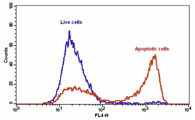
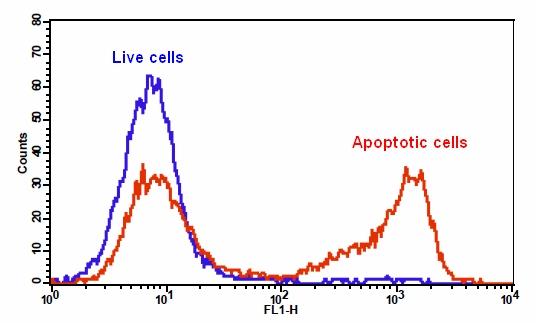
图1.在Jurkat细胞中检测Apopxin Green和磷脂酰丝氨酸的结合活性。 将Jurkat细胞在37℃,5%CO2培养箱中在没有(蓝色)或20μM喜树碱(红色)的情况下处理5小时,然后用Apopxin Green染色15分钟。使用FL1通道,用FACSCalibur(Becton Dickinson)流式细胞仪测量Apopxin Green的荧光强度。


参考文献

Combined Treatments of Magnetic Intra-Lysosomal Hyperthermia with Doxorubicin Promotes Synergistic Anti-Tumoral Activity.
Authors: D Hajj Diab El, P Clerc, N Serhan, D Fourmy, V Gigoux
Journal: Nanomaterials (Basel, Switzerland) (2018)


相关产品

产品名称 货号
Cell Meter 磷脂酰丝氨酸细胞凋亡检测试剂盒 红色荧光 适合于流式细胞检测 Cat#22832
Cell Meter 磷脂酰丝氨酸凋亡检测试剂盒 红色荧光,适合微孔板检测 Cat#22792
Cell Meter 磷脂酰丝氨酸凋亡检测试剂盒 绿色荧光,适合微孔板检测 Cat#22791

Cell Meter 磷脂酰丝氨酸细胞凋亡检测试剂盒 红色荧光 适合于流式细胞检测-AAT Bioquest荧光染料

上海金畔生物科技有限公司代理AAT Bioquest荧光染料全线产品,欢迎访问AAT Bioquest荧光染料官网了解更多信息。
Cell Meter 磷脂酰丝氨酸细胞凋亡检测试剂盒 红色荧光 适合于流式细胞检测 价格 2823
产品规格

100 Tests

产品货号

Cell Meter 磷脂酰丝氨酸细胞凋亡检测试剂盒 红色荧光 适合于流式细胞检测

产品参数
Ex (nm) Em (nm)
分子量 溶剂
存储条件
产品概述

我们的Cell Meter 检测试剂盒是一套用于检测细胞生存力的工具。有多种参数可用于检测细胞活力。 该特定试剂盒旨在通过测量磷脂酰丝氨酸(PS)的转运来检测细胞凋亡。在细胞凋亡中,PS转移到质膜的外部小叶。磷脂酰丝氨酸在细胞表面的出现是细胞凋亡初始/中间阶段的普遍指标,可以在观察形态变化之前进行检测。该试剂盒中使用的我们专有的Apopxin PS探针是基于小分子的PS探针。 与膜PS结合后具有红色荧光。 使用流式细胞仪在APC通道(红色荧光)优化了此特定的检测试剂盒,以检测细胞凋亡。金畔生物是AAT Bioquest的中国代理商,为您提供优质的Cell Meter 磷脂酰丝氨酸细胞凋亡检测试剂盒。


适用仪器


流式细胞仪  
Ex: 640 nm
Em: 660/20   nm 
通道: APC 通道
实验方案

样品实验方案

简要概述

  1. 用测试化合物制备细胞(200 µL /样品)
  2. 添加Apopxin 深红色测定溶液
  3. 在室温下孵育30-60分钟
  4. 使用具有FL4通道(Ex / Em = 647/660 nm的流式细胞仪)分析细胞


实验步骤

1.用测试化合物处理细胞所需的时间(喜树碱处理过的Jurkat细胞需要4-6小时)以凋亡。 

2.离心细胞以获得1-5×10⁵细胞/管。

3.将细胞重悬于200 µL分析缓冲液(组分B)中。

4.向细胞中加入2 µL Apopxin 深红色溶液(组分A)。

5.避光保存,于室温下孵育30至60分钟。

6.可选:在使用流式细胞仪分析细胞之前,添加200至300 µL分析缓冲液(组分B)以增加体积。

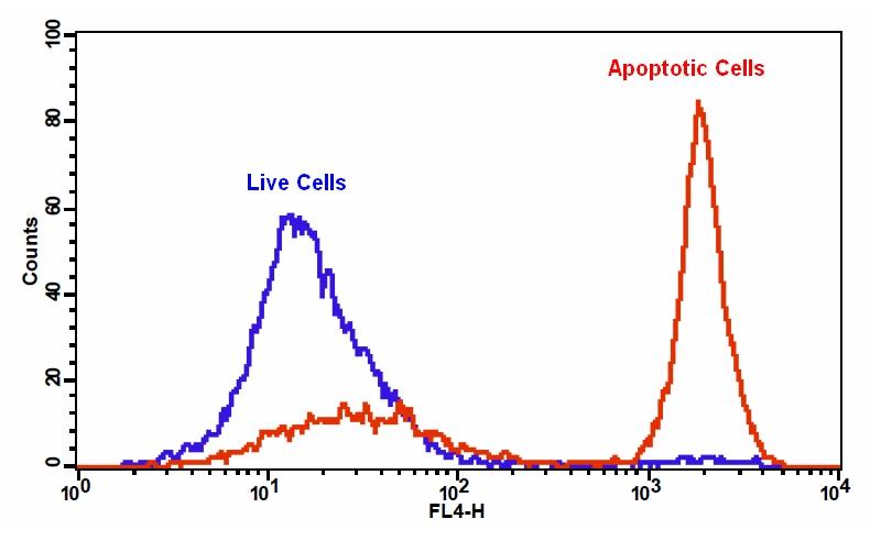
7.使用带有FL4通道的流式细胞仪(Ex / Em = 647/660 nm)检测荧光强度。


参考文献

Apoptosis of human Burkitt’s lymphoma cells induced by 2-N,N-diethylaminocarbonyloxymethyl-1-diphenylmethyl-4-(3,4,5-trimethoxybe nzoyl) piperazine hydrochloride (PMS-1077)
Authors: Wang WD, Xu XM, Chen Y, Jiang P, Dong CZ, Wang Q.
Journal: Arch Pharm Res (2009): 1727

Detection of apoptosis based on the interaction between annexin V and phosphatidylserine
Authors: Liu T, Zhu W, Yang X, Chen L, Yang R, Hua Z, Li G.
Journal: Anal Chem (2009): 2410

Dynamic analysis of apoptosis using cyanine SYTO probes: from classical to microfluidic cytometry
Authors: Wlodkowic D, Skommer J, Faley S, Darzynkiewicz Z, Cooper JM.
Journal: Exp Cell Res (2009): 1706

Eurycomanone induce apoptosis in HepG2 cells via up-regulation of p53
Authors: Zakaria Y, Rahmat A, Pihie AH, Abdullah NR, Houghton PJ.
Journal: Cancer Cell Int (2009): 16

Evaluation of cell surface expression of phosphatidylserine in ovarian carcinoma effusions using the annexin-V/7-AAD assay: clinical relevance and comparison with other apoptosis parameters
Authors: Dong HP, Holth A, Kleinberg L, Ruud MG, Elstr and MB, Trope CG, Davidson B, Risberg B.
Journal: Am J Clin Pathol (2009): 756

Glycogen synthase kinase-3 and Omi/HtrA2 induce annexin A2 cleavage followed by cell cycle inhibition and apoptosis
Authors: Wang CY, Lin YS, Su WC, Chen CL, Lin CF.
Journal: Mol Biol Cell (2009): 4153

Gold fluorescent annexin A5 as a novel apoptosis detection tool
Authors: Kurschus FC, Pal PP, Baumler P, Jenne DE, Wiltschi B, Budisa N.
Journal: Cytometry A (2009): 626

Induction of apoptosis in sonoporation and ultrasonic gene transfer
Authors: Miller DL, Dou C.
Journal: Ultrasound Med Biol (2009): 144

Mobilization of lysosomal calcium regulates the externalization of phosphatidylserine during apoptosis
Authors: Mirnikjoo B, Balasubramanian K, Schroit AJ.
Journal: J Biol Chem (2009): 6918

Peptidic targeting of phosphatidylserine for the MRI detection of apoptosis in atherosclerotic plaques
Authors: Burtea C, Laurent S, Lancelot E, Ballet S, Murariu O, Rousseaux O, Port M, V and er Elst L, Corot C, Muller RN.
Journal: Mol Pharm (2009): 1903

Cell Meter 荧光法细胞周期检测试剂盒 绿色荧光 适合于流式细胞检测-AAT Bioquest荧光染料

上海金畔生物科技有限公司代理AAT Bioquest荧光染料全线产品,欢迎访问AAT Bioquest荧光染料官网了解更多信息。
Cell Meter 荧光法细胞周期检测试剂盒 绿色荧光 适合于流式细胞检测 价格 2823
产品规格

100 Tests

产品货号

Cell Meter 荧光法细胞周期检测试剂盒 绿色荧光 适合于流式细胞检测

产品参数
Ex (nm) 503 Em (nm) 527
分子量 溶剂
存储条件
产品概述

Cell Meter 荧光法细胞周期检测试剂盒是美国AAT Bioquest生产的用于检测细胞周期的试剂盒,Cell Meter检测试剂盒是一类用于检测细胞功能的系列工具,包括细胞活性、细胞毒性、细胞凋亡、细胞膜电位以及细胞周期等方面的指标。每种检测方案均能提供不同荧光颜色的检测方案。这些高效的检测方案为从多角度研究细胞功能活动提供了一种十分有效的方法。在正常细胞中,DNA密度的改变与细胞生长、分化、休眠等细胞功能相关。细胞周期通过许多不同的细胞周期调控因子相互作用来调节。这些调控因子,可以与DNA结合,开启或关闭那些能引起细胞分裂的蛋白质合成。在这个调控级联反应中,任何一步的错误都会引起细胞非正常地增殖,这是一种潜在的病理状态,例如肿瘤的形成。活细胞研究的潜在应用是对细胞DNA含量的测定和细胞周期进行检测,为了对细胞的生长模式的变化进行检测,对细胞凋亡进行监测,对肿瘤细胞活动和抑制剂基因机制进行研究。Cell Meter荧光法细胞周期检测试剂盒 *绿色荧光 适合于流式细胞检测* 是设计用来检测细胞周期进程和增殖的,在活细胞、经通透性处理的细胞和固定化细胞中使用我们独特的Nuclear Green CCS1对细胞周期进程和增殖进行监测。在所给样品的不同细胞中,有G0/G1, S 和G2/M 期以及细胞凋亡之前的亚-G1期等,可以通过流式细胞仪检测。经过Nuclear Green CC1染色的细胞可以通过流式细胞仪在Ex/Em = 490 nm/520 nm(FL1 通道)进行检测。金畔生物是AAT Bioquest的中国代理商,为您提供优质的Cell Meter 荧光法细胞周期检测试剂盒。

点击查看光谱 


适用仪器


流式细胞仪  
Ex: 488 nm
Em: 530/30  nm 
通道: FITC 通道
实验方案

样品实验方案

简要概述

用测试化合物制备细胞,密度为5×10⁵至1×10⁶个细胞/ mL
将2.5μL200XNuclear Green CCS1加入0.5 mL细胞溶液中
在37°C,5%CO2培养箱中孵育30-60分钟
使用FL1通道用流式细胞仪分析细胞((Ex / Em = 490/525 nm)


操作步骤

1.对于每个样品,将细胞制备在0.5 mL温热培养基或您选择的缓冲液中,密度为5×10⁵至1×10⁶个细胞/ mL。注意:应对每个细胞系进行单独评估,以确定诱导细胞凋亡的佳细胞密度。

2.用测试化合物处理细胞一段所需的时间以诱导细胞凋亡或其他细胞周期功能。

3.将2.5μL的200X Nuclear Green CCS1(组分A)加入处理过的细胞中。

4.将细胞在37°C,5%CO2培养箱中孵育30至60分钟。注意:对于粘附细胞,用0.5 mM EDTA轻轻提起细胞以保持细胞完整,并在与Nuclear Green CCS1孵育之前用含血清的培养基洗涤细胞一次。适当的孵育时间取决于所用的单个细胞类型和细胞浓度。优化每个实验的孵育时间。由于Nuclear Green CCS1是细胞可渗透的,因此不必在DNA染色之前固定细胞。

5.可选:将细胞以1000 rpm离心4分钟,然后将细胞重新悬浮于0.5 mL测定缓冲液(组分B)或您选择的缓冲液中。

6.使用具有FL1通道的流式细胞仪(Ex / Em = 490 / 525nm)监测荧光强度。


数据分析

Cell Meter 荧光法细胞周期检测试剂盒 绿色荧光 适合于流式细胞检测

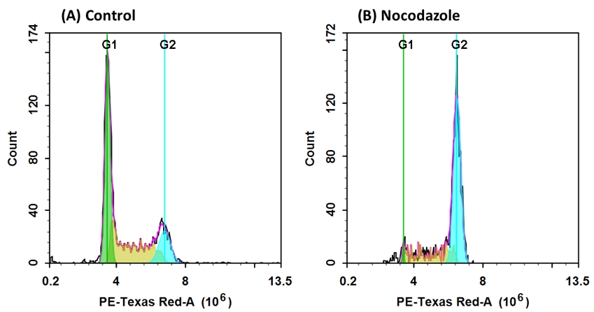
图1生长和诺考达唑处理的Jurkat细胞中的DNA谱。 Jurkat细胞在37℃,5%CO2培养箱中不用(A)或用100ng / ml诺考达唑(B)处理24小时,然后用Nuclear Green CCS1染色30分钟。 使用具有FITC通道的ACEA NovoCyte流式细胞仪测量Nuclear Green CCS1的荧光强度。 在生长Jurkat细胞(A)中,用Nuclear Green CCS1染色的核显示出G1,S和G2期。 在Nocodazole处理G2阻滞细胞(B)后,G2细胞频率急剧增加,G1期,S期频率明显下降。


参考文献

Cell cycle synchronization of Escherichia coli using the stringent response, with fluorescence labeling assays for DNA content and replication
Authors: Ferullo DJ, Cooper DL, Moore HR, Lovett ST.
Journal: Methods (2009): 8

DNA replication, cell cycle progression and the targeted gene repair reaction
Authors: Engstrom JU, Kmiec EB.
Journal: Cell Cycle (2008): 1402

Morin inhibits the growth of human leukemia HL-60 cells via cell cycle arrest and induction of apoptosis through mitochondria dependent pathway
Authors: Kuo HM, Chang LS, Lin YL, Lu HF, Yang JS, Lee JH, Chung JG.
Journal: Anticancer Res (2007): 395

Direct control of cell cycle gene expression by proto-oncogene product ACTR, and its autoregulation underlies its transforming activity
Authors: Louie MC, Revenko AS, Zou JX, Yao J, Chen HW.
Journal: Mol Cell Biol (2006): 3810

Cell cycle markers for live cell analyses
Authors: Easwaran HP, Leonhardt H, Cardoso MC.
Journal: Cell Cycle (2005): 453

Dynamic relocalization of hOGG1 during the cell cycle is disrupted in cells harbouring the hOGG1-Cys326 polymorphic variant
Authors: Luna L, Rolseth V, Hildrestrand GA, Otterlei M, Dantzer F, Bjoras M, Seeberg E.
Journal: Nucleic Acids Res (2005): 1813

Dynamics of relative chromosome position during the cell cycle
Authors: Essers J, van Cappellen WA, Theil AF, van Drunen E, Jaspers NG, Hoeijmakers JH, Wyman C, Vermeulen W, Kanaar R.
Journal: Mol Biol Cell (2005): 769

Cell cycle regulation of the murine 8-oxoguanine DNA glycosylase (mOGG1): mOGG1 associates with microtubules during interphase and mitosis
Authors: Conlon KA, Zharkov DO, Berrios M.
Journal: DNA Repair (Amst) (2004): 1601

Description of a flow cytometry approach based on SYBR-14 staining for the assessment of DNA content, cell cycle analysis, and sorting of living normal and neoplastic cells
Authors: Nunez R, Garay N, Villafane C, Bruno A, Lindgren V.
Journal: Exp Mol Pathol (2004): 29

Differential roles of STAT1alpha and STAT1beta in fludarabine-induced cell cycle arrest and apoptosis in human B cells
Authors: Baran-Marszak F, Feuillard J, Najjar I, Le Clorennec C, Bechet JM, Dusanter-Fourt I, Bornkamm GW, Raphael M, Fagard R.
Journal: Blood (2004): 2475


相关产品

产品名称 货号
Cell Meter 荧光法活细胞周期检测试剂盒 红色荧光 适用于流式细胞仪 Cat#22860
Cell Meter 荧光法细胞周期检测试剂盒 405nm激发 Cat#22845
Cell Meter 荧光法细胞周期检测试剂盒 红色荧光 适合于流式细胞检测 Cat#22842

Cell Meter 荧光法细胞周期检测试剂盒 红色荧光 适合于流式细胞检测-AAT Bioquest荧光染料

上海金畔生物科技有限公司代理AAT Bioquest荧光染料全线产品,欢迎访问AAT Bioquest荧光染料官网了解更多信息。
Cell Meter 荧光法细胞周期检测试剂盒 红色荧光 适合于流式细胞检测 价格 2823
产品规格

100 Tests

产品货号

Cell Meter 荧光法细胞周期检测试剂盒 红色荧光 适合于流式细胞检测

产品参数
Ex (nm) 537 Em (nm) 618
分子量 溶剂
存储条件
产品概述

我们的Cell Meter 检测试剂盒是一套用于检测细胞活力和增殖的工具。有多种参数可用于监测细胞活力和增殖。在正常细胞中,DNA密度根据细胞的生长,分裂,静止执行其正常功能而变化。细胞周期的进程受各种细胞周期调节因子之间复杂的相互作用控制。这些调节剂转录因子,该转录因子与DNA结合并开启或关闭导致细胞分裂的蛋白质的产生。这种调节级联中的任何失误都会导致异常细胞增殖,这是许多病理状况(例如肿瘤形成)的基础。活细胞研究的潜在应用是确定细胞DNA含量和细胞周期分布,以检测生长模式的变化,检测凋亡,评估肿瘤细胞的行为和抑制基因的机制。该特定试剂盒旨在使用Nuclear Red CCS1(固定细胞中的细胞周期染色剂)检测细胞周期进程和增殖。染料穿过透化的膜并插入细胞DNA中。试剂盒中提供的RNase降解RNA后,Nuclear Red CCS1的信号强度与DNA含量成正比。可以使用流式细胞仪(FL2通道)检测给定样品中处于G0 / G1,S和G2 / M期的细胞百分比,以及处于凋亡之前的sub-G1期的细胞百分比。金畔生物是AAT Bioquest的中国代理商,为您提供优质的Cell Meter 荧光法细胞周期检测试剂盒。

点击查看光谱


适用仪器


流式细胞仪  
Ex: 488 nm
Em: 610/20   nm 
通道: PE-Texas Red 通道
实验方案

样品实验方案

简要概述

  1. 用5×10⁵至1×10⁶cell/ mL的密度制备含测试化合物的细胞
  2. 用70%乙醇固定细胞
  3. 将0.5 µL 100X Nuclear Red CCS1和5 µL RNase A加入0.5 mL细胞溶液中
  4. 在室温下孵育30-60分钟
  5. 使用带有FL2通道的流式细胞仪分析细胞


实验步骤

1.用测试化合物处理细胞,以诱导凋亡或其他细胞周期功能。

2.对于每个样品,用0.5 mL PBS制备细胞,密度为5×10⁵至1×10⁶细胞/ mL。

2.1对于贴壁细胞:将细胞用胰蛋白酶消化,悬浮在10%FBS培养基中,离心(1000 rpm,5分钟),然后将沉淀重悬于PBS中。

2.2对于悬浮细胞:将细胞离心(1000 rpm,5分钟),并将沉淀物悬浮在PBS(1 mL)中。 注意:应单独评估每种细胞系,以确定诱导凋亡的细胞密度。

3.用70%乙醇固定细胞:

3.1将0.5 mL细胞悬液吸移至1.2 mL无水乙醇中(浓度约为70%)。

3.2在冰上孵育细胞至少2小时(或在-20°C过夜)。在染色之前,细胞可以在-20°C下保存2年。注意:在用单克隆抗体染色细胞表面抗原后,通常将乙醇用于固定,而在用单克隆抗体染色细胞内抗原后,通常将甲醇用于固定。在此过程中,将固定并分析整个细胞。由于仍然存在整个细胞团,因此通常包括使用RNase来任何双链RNA。尽管已经对整个细胞进行了分析,但尝试检测与DNA结合的某些细胞内抗原的尝试可能会失败,因为蛋白质会从透化的细胞中漏出(例如绿色荧光蛋白)。在这些情况下,在酒精固定之前,用PBS中的1%低聚甲醛进行短暂的预固定(在4-6°C下10分钟),将有助于将蛋白质保留在细胞中。

4.用Nuclear Red CCS1染色细胞:

4.1以1000 rpm的速度沉淀细胞5分钟,并用冷PBS至少洗涤一次。

4.2将沉淀物悬浮在0.5 mL测定缓冲液(组分C)中。

4.3加入5 µL 100X Nuclear Red CCS1(组分A)和5 µL 100X RNase A(组分B)。

4.4在室温下孵育细胞30至60分钟。 注意:适当的孵育时间取决于所用的单个细胞类型和细胞浓度。 优化每个实验的孵育时间。

4.5可选:将细胞以1000 rpm离心5分钟,然后将细胞重悬于0.5 mL检测缓冲液(组分B)或自备缓冲液中。

4.6使用FL2通道(Ex / Em = 490/620 nm),通过流式细胞仪检测荧光强度。 


参考文献 

Cell cycle synchronization of Escherichia coli using the stringent response, with fluorescence labeling assays for DNA content and replication
Authors: Ferullo DJ, Cooper DL, Moore HR, Lovett ST.
Journal: Methods (2009): 8

DNA replication, cell cycle progression and the targeted gene repair reaction
Authors: Engstrom JU, Kmiec EB.
Journal: Cell Cycle (2008): 1402

Morin inhibits the growth of human leukemia HL-60 cells via cell cycle arrest and induction of apoptosis through mitochondria dependent pathway
Authors: Kuo HM, Chang LS, Lin YL, Lu HF, Yang JS, Lee JH, Chung JG.
Journal: Anticancer Res (2007): 395

Direct control of cell cycle gene expression by proto-oncogene product ACTR, and its autoregulation underlies its transforming activity
Authors: Louie MC, Revenko AS, Zou JX, Yao J, Chen HW.
Journal: Mol Cell Biol (2006): 3810

Cell cycle markers for live cell analyses
Authors: Easwaran HP, Leonhardt H, Cardoso MC.
Journal: Cell Cycle (2005): 453

Dynamic relocalization of hOGG1 during the cell cycle is disrupted in cells harbouring the hOGG1-Cys326 polymorphic variant
Authors: Luna L, Rolseth V, Hildrestr and GA, Otterlei M, Dantzer F, Bjoras M, Seeberg E.
Journal: Nucleic Acids Res (2005): 1813

Dynamics of relative chromosome position during the cell cycle
Authors: Essers J, van Cappellen WA, Theil AF, van Drunen E, Jaspers NG, Hoeijmakers JH, Wyman C, Vermeulen W, Kanaar R.
Journal: Mol Biol Cell (2005): 769

Cell cycle regulation of the murine 8-oxoguanine DNA glycosylase (mOGG1): mOGG1 associates with microtubules during interphase and mitosis
Authors: Conlon KA, Zharkov DO, Berrios M.
Journal: DNA Repair (Amst) (2004): 1601

Description of a flow cytometry approach based on SYBR-14 staining for the assessment of DNA content, cell cycle analysis, and sorting of living normal and neoplastic cells
Authors: Nunez R, Garay N, Villafane C, Bruno A, Lindgren V.
Journal: Exp Mol Pathol (2004): 29

Differential roles of STAT1alpha and STAT1beta in fludarabine-induced cell cycle arrest and apoptosis in human B cells
Authors: Baran-Marszak F, Feuillard J, Najjar I, Le Clorennec C, Bechet JM, Dusanter-Fourt I, Bornkamm GW, Raphael M, Fagard R.
Journal: Blood (2004): 2475

Screen Quest Fluo-8 去除培养基钙检测试剂盒*适合于难检测细胞系*-AAT Bioquest荧光染料

上海金畔生物科技有限公司代理AAT Bioquest荧光染料全线产品,欢迎访问AAT Bioquest荧光染料官网了解更多信息。
Screen Quest Fluo-8 去除培养基钙检测试剂盒*适合于难检测细胞系*价格 1386
产品规格

1 Plate

产品货号

Screen Quest Fluo-8 去除培养基钙检测试剂盒*适合于难检测细胞系*

产品参数
Ex (nm) 495 Em (nm) 516
分子量 溶剂
存储条件 在零下15度以下保存, 避免光照
产品概述

Screen Quest Fluo-8钙检测试剂盒是美国AAT Bioquest生产的钙检测试剂盒,钙通量测定是用于筛选G蛋白偶联受体(GPCR)的药物发现中的优选方法。 Screen Quest Fluo-8 NW钙测定试剂盒提供均匀的荧光测定,用于检测细胞内钙动员。表达感兴趣的GPCR的细胞通过钙预先加载我们专有的Fluo-8 NW,其可以穿过细胞膜。 Fluo-8 NW是可用于HTS筛选的亮钙指示剂。一旦进入细胞内,Fluo-8 NW的亲脂性阻断基团被非特异性细胞酯酶切割,产生带负电荷的荧光染料,其停留在细胞内,并且在与钙结合时其荧光大大增强。当用筛选化合物刺激细胞时,该受体表明细胞内钙的释放,这极大地增加了Fluo-8 NW的荧光。其长波长,高灵敏度和100-250倍荧光增加的特性(当它与钙形成复合物时)使Fluo-8 NW成为测量细胞钙的理想指标。这个Screen Quest Fluo-8 NW钙测定试剂盒提供了一种优化的检测方法,用于监测G蛋白偶联受体(GPCR)和钙通道。该测定可以以方便的96孔或384孔微量滴定板形式进行,并且易于适应自动化。金畔生物是AAT Bioquest的中国代理商,为您提供优质的Screen Quest Fluo-8 去除培养基钙检测试剂盒*适合于难检测细胞系*。 

点击查看光谱


适用仪器


荧光酶标仪  
Ex: 490 nm
Em: 525 nm
Cutoff: 515 nm
推荐孔板: 黑色透明底板
读取模式: 底部读取/分液处理


其他可用仪器  
ArrayScan、FDSS、FLIPR、FlexStation、IN Cell Analyzer、NOVOStar、ViewLux  
实验方案

样品实验方案

简要概述

1.准备细胞
2.去除生长培养基
3.添加Fluo-8 NW染料工作溶液
4.在室温下孵育1小时
5.监测Ex / Em = 490 / 525nm处的荧光强度


溶液配制

1.储存溶液配制

除非另有说明,否则所有未使用的储备溶液应分成一次性等分试样,并在制备后储存在-20°C。 避免反复冻融循环。
1.1 Fluo-8 NW配制:
对于Cat No.36307,将10μLDMSO加入Fluo-8 NW(组分A)中,并充分混合。
对于Cat No.36308和36309,将100μLDMSO加入Fluo-8 NW(组分A)中,并充分混合。 注意:10μLFluo-8 NW原液足以容纳1个平板。

1.2分析缓冲液储备液(1X):
对于Cat No.36307和36308,将9mL HHBS(组分C)加入10X Pluronic F127 Plus(1mL,组分B)中并充分混合。
对于Cat No.36309,将整瓶10XPluronic®F127Plus(10 mL,组分B)加入90 mL HHBS缓冲液(不包括在试剂盒中)中并充分混合。 注意:对于一个平板,10 mL的1X分析缓冲液就足够了。


2.工作溶液配制

将10μLFluo-8 NW DMSO储备溶液加入10mL 1X测定缓冲液中并充分混合。 该工作溶液在室温下稳定至少2小时。


操作步骤

1.有关细胞样品制备的指南,请点击查看。

2.从细胞板上除去生长培养基。注意:重要的是去除生长培养基,以大限度地减少背景荧光和化合物对血清或培养基的干扰。注意:或者,在含有0.5%至1%FBS的生长培养基中培养细胞,以避免培养基去除步骤。在这种情况下,需要在HHBS缓冲液中加入2X染料。 [我们为那些在生长培养基中使用0.5%至1%FBS以避免培养基去除步骤的人提供2个独立的无洗涤钙测定试剂盒(目录号36315和目录号36316)。

3.将100μL/孔(96孔板)或25μL/孔(384孔板)的Fluo-8NW染料工作溶液加入到细胞板中。

4.将染料加载板在细胞培养箱中孵育30分钟,然后在室温下将板孵育另外30分钟。注意:如果测定需要37°C,立即进行实验,无需进一步室温孵育。注意:如果细胞在室温下运行时间较长,可在室温下孵育细胞培养板1-2小时(建议培养时间不超过2小时。)

5.用HHBS或所需缓冲液制备复合板。

6.通过监测Ex / Em = 490 / 525nm处的荧光强度来进行钙通量测定。注意:在实验之前运行信号测试很重要。不同的仪器有自己的强度范围。将信号测试强度调整到大强度计数的10%至15%的水平。例如,FLIPR-384的大荧光强度计数为65,000,因此应调整仪器设置以使其信号测试强度在7,000到10,000左右。


数据分析

        从空白标准孔获得的读数(RFU(Max-Min))用作阴性对照。 从其他标准的读数中减去该值,以获得基线校正值。 然后,绘制标准读数以获得标准曲线和方程。 该等式可用于计算卡巴胆碱剂量样品。 

Screen Quest Fluo-8 去除培养基钙检测试剂盒*适合于难检测细胞系*

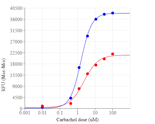
图1.使用Screen Quest Fluo-8 NW Assay Kit和Fluo-4 NW Assay Kit在HEK-293细胞中测量Carbachol剂量反应。 将HEK-293细胞以40,000个细胞/100μL/孔接种在Costar黑壁/透明底96孔板中过夜。 除去生长培养基,使用Screen Quest Fluo 8-NW钙测定试剂盒或Fluo-4 NW试剂盒(根据制造商的说明书)将细胞与100μL染料上样溶液一起在室温下孵育1小时 温度。 通过NOVOstar(BMG Labtech)添加卡巴胆碱(25μL/孔)以达到指示的浓度。 Fluo8 NW的EC50约为1.2μM。


参考文献

2-OMe-lysophosphatidylcholine analogues are GPR119 ligands and activate insulin secretion from βTC-3 pancreatic cells: Evaluation of structure-dependent biological activity
Authors: Anna Drzazga, Agata Sowińska, Agnieszka Krzemińska, Andrzej Okruszek, Piotr Paneth, Maria Koziolkiewicz, Edyta Gendaszewska-Darmach
Journal: Biochimica et Biophysica Acta (BBA)-Molecular and Cell Biology of Lipids (2017)

A new electro-optical approach for conductance measurement: an assay for the study of drugs acting on ligand-gated ion channels
Authors: A Menegon, S Pitassi, N Mazzocchi, L Redaelli, R Rizzetto, JF Rolland, C Poli, M Imberti, A Lanati, F Grohovaz
Journal: Scientific Reports (2017)

Altered spontaneous calcium signaling of in situ chondrocytes in human osteoarthritic cartilage
Authors: Xiaoyuan Gong, Wenbin Xie, Bin Wang, Lingchuan Gu, Fuyou Wang, Xiang Ren, Cheng Chen, Liu Yang
Journal: Scientific reports (2017): 17093

Analysis of Ca2+ response of osteocyte network by three-dimensional time-lapse imaging in living bone
Authors: Tomoyo Tanaka, Mitsuhiro Hoshijima, Junko Sunaga, Takashi Nishida, Mana Hashimoto, Naoya Odagaki, Ryuta Osumi, Taiji Aadachi, Hiroshi Kamioka
Journal: Journal of Bone and Mineral Metabolism (2017): 1–10

Aryl-and alkyl-phosphorus-containing flame retardants induced mitochondrial impairment and cell death in Chinese hamster ovary (CHO-k1) cells
Authors: Chao Huang, Na Li, Shengwu Yuan, Xiaoya Ji, Mei Ma, Kaifeng Rao, Zijian Wang
Journal: Environmental Pollution (2017): 775–786

Bystander effects elicited by single-cell photo-oxidative blue-light stimulation in retinal pigment epithelium cell networks
Authors: Masaaki Ishii, Bärbel Rohrer
Journal: Cell Death Discovery (2017): 16071

Ca 2+ signals initiate at immobile IP 3 receptors adjacent to ER-plasma membrane junctions
Authors: Nagendra Babu Thillaiappan, Alap P Chavda, Stephen C Tovey, David L Prole, Colin W Taylor
Journal: Nature Communications (2017): 1505

Cells smell on a CMOS: A portable odorant detection system using cell-laden collagen pillars
Authors: Yusuke Hirata, Yuya Morimoto, Eunryel Nam, Shotaro Yoshida, Shoji Takeuchi
Journal: (2017): 13–16

Ex vivo replication of phenotypic functions of osteocytes through biomimetic 3D bone tissue construction
Authors: Qiaoling Sun, Saba Choudhary, Ciaran Mannion, Yair Kissin, Jenny Zilberberg, Woo Y Lee
Journal: Bone (2017)

High Glucose Enhances Isoflurane-Induced Neurotoxicity by Regulating TRPC-Dependent Calcium Influx
Authors: ZhongJie Liu, ChangQing Ma, Wei Zhao, QingGuo Zhang, Rui Xu, HongFei Zhang, HongYi Lei, ShiYuan Xu
Journal: Neurochemical Research (2017): 1–14


相关产品

产品名称 货号
Screen Quest Calbryte 520 免丙磺舒和免洗钙检测试剂盒 Cat#36317
Screen Quest 免洗Fluo-8钙检测试剂盒 Cat#36314