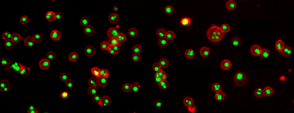
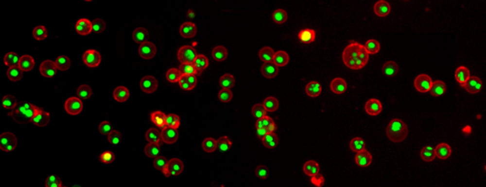
BD 559763-细胞凋亡试剂盒(PE和7-ADD标记)PE Annexin V Apoptosis Detectio

【简单介绍】

NameAnnexin V : PE Apoptosis Detection Kit IContentsAnnexin V-PE, 7-AAD, and Annexin V Binding BufferSize100 TestsRegulatory Status RUO

*,现货*格优惠,咨询

【详细说明】

 Technical Data Sheet

PE Annexin V Apoptosis Detection Kit I

Product Information

Material Number: 559763

Component: 51-66121E

Description: 10X Annexin V Binding Buffer

Size: 50 ml (1 ea)

Storage Buffer: Aqueous buffered solution containing no preservative.

Component: 51-68981E

Description: 7-AAD

Size: 2.0 ml (1 ea)

Vol. per Test: 5 μl

Storage Buffer: Aqueous buffered solution containing fetal bovine serum and ≤0.09% sodium

azide.

Component: 51-65875X

Description: PE Annexin V

Size: 0.5 ml (1 ea)

Vol. per Test: 5 μl

Storage Buffer: Aqueous buffered solution containing BSA and ≤0.09% sodium azide.

Description

Apoptosis is a normal physiologic process which occurs during embryonic development as well as in maintenence of tissue homeostasis. The

apoptotic program is characterized by certain morphologic features, including loss of plasma membrane asymmetry and attachment,

condensation of the cytoplasm and nucleus, and internucleosomal cleavage of DNA. Loss of plasma membrane is one of the earliest features.

In apoptotic cells, the membrane phospholipid phosphatidylserine (PS) is translocated from the inner to the outer leaflet of the plasma

membrane, thereby exposing PS to the external cellular environment. Annexin V is a 35-36 kDa Ca2+ dependent phospholipid-binding

protein that has a high affinity for PS, and binds to cells with exposed PS. Annexin V may be conjugated to fluorochromes including

Phycoerythrin (PE). This format retains its high affinity for PS and thus serves as a sensitive probe for flow cytometric analysis of cells that are

undergoing apoptosis. Since externalization of PS occurs in the earlier stages of apoptosis, PE Annexin V staining can identify apoptosis at an

earlier stage than assays based on nuclear changes such as DNA fragmentation.

PE Annexin V staining precedes the loss of membrane integrity which accompanies the latest stages of cell death resulting from either

apoptotic or necrotic processes. Therefore, staining with PE Annexin V is typically used in conjunction with a vital dye such as

7-Amino-Actinomycin (7-AAD) to allow the investigator to identify early apoptotic cells (7-AAD negative, PE Annexin V positive). Viable

cells with intact membranes exclude 7-AAD, wheras the membranes of dead and damaged cells are permeable to 7-AAD. For example, cells

that are considered viable are PE Annexin V and 7-AAD negative; cells that are in early apoptosis are PE Annexin V positive and 7-AAD

negative; and cells that are in late apoptosis or already dead are are both PE Annexin V and 7-AAD positive. This assay does not distinguish

between cells that have undergone apoptotic death versus those that have died as a result of a necrotic pathway because in either case, the dead

cells will stain with both PE Annexin V and 7-AAD. However, when apoptosis is measured over time, cells can be often tracked from PE

Annexin V and 7-AAD negative (viable, or no measurable apoptosis), to PE Annexin V positive and 7-AAD negative (early apoptosis,

membrane integrity is present) and finally to PE Annexin V and 7-AAD positive (end stage apoptosis and death). The movement of cells

through these three stages suggests apoptosis. In contrast, a single observation indicating that cells are both PE Annexin V and 7-AAD

positive, in of itself, reveals less information about the process by which the cells underwent their demise.

559763 Rev. 8 Page 1 of 3

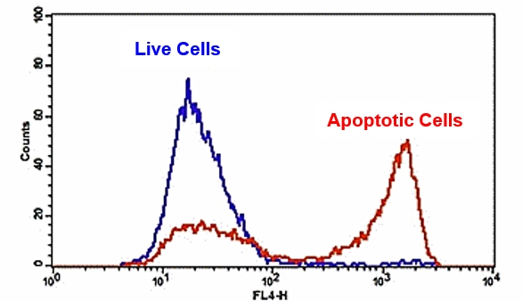
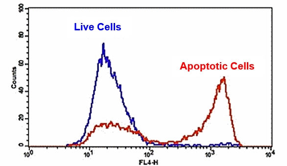
Flow Cytometric Analysis of PE Annexin V staining. Jurkat cells

(Human T-cell leukemia; ATCC TIB-152) were left untreated (top

panels) or treated for 4 hours with 4 μM Camptothecin (bottom

panels). Cells were incubated with PE Annexin V in a buffer

containing 7-Amino-Actinomycin (7-AAD) and analyzed by flow

cytometry. Untreated cells were primarily PE Annexin V and 7-AAD

negative, indicating that they were viable and not undergoing

apoptosis. After a 4 hour treatment (bottom panels), there were

primarily two populations of cells: Cells that were viable and not

undergoing apoptosis (PE Annexin V and 7-AAD negative); cells

undergoing apoptosis (PE Annexin V positive and 7-AAD negative).

A minor population of cells were observed to be PE Annexin V and

7-AAD positive, indicating that they were in end stage apoptosis or

already dead.

Preparation and Storage

Store undiluted at 4°C and protected from prolonged exposure to light. Do not freeze.

Application Notes

Application

Flow cytometry Routinely Tested

Recommended Assay Procedure:

PE Annexin V is a sensitive probe for identifying apoptotic cells, binding to negatively charged phospholipid surfaces (Kd of ~5 x 10^-2) with a

higher affinity for phosphatidylserine (PS) than most other phospholipids. PE Annexin V binding is calcium dependent and defined calcium and

salt concentrations are required for optimal staining as described in the PE Annexin V Staining Protocol. Investigators should note that PE

Annexin V flow cytometric analysis on adherent cell types (e.g HeLa, NIH 3T3, etc.) is not routinely tested as specific membrane damage

may occur during cell detachment or harvesting. Methods for utilizing Annexin V for flow cytometry on adherent cell types, however,

have been previously reported (Casiola-Rosen et al. and van Engelend et al.).

INDUCTION OF APOPTOSIS BY CAMPTOTHECIN

The following protocol is provided as an illustration on how PE Annexin V may be used on a cell line (Jurkat).

Materials

1. Prepare Camptothecin stock solution (Sigma-Aldrich Cat. No. C-9911): 1 mM in DMSO.

2. Jurkat T cells (ATCC TIB-152).

Procedure

1. Add Camptothecin (final conc. 4-6 μM) to 1 x 10^6 Jurkat cells.

2. Incubate the cells for 4-6 hr at 37°C.

3. Proceed with the PE Annexin V Staining Protocol to measure apoptosis.

PE ANNEXIN V STAINING PROTOCOL

PE Annexin V is used to quantitatively determine the percentage of cells within a population that are actively undergoing apoptosis. It relies on

the property of cells to lose membrane asymmetry in the early phases of apoptosis. In apoptotic cells, the membrane phospholipid

phosphatidylserine (PS) is translocated from the inner leaflet of the plasma membrane to the outer leaflet, thereby exposing PS to the external

environment. Annexin V is a calcium-dependent phospholipid-binding protein that has a high affinity for PS, and is useful for identifying

apoptotic cells with exposed PS. 7-Amino-Actinomycin (7-AAD) is a standard flow cytometric viability probe and is used to distinguish viable

from nonviable cells. Viable cells with intact membranes exclude 7-AAD, whereas the membranes of dead and damaged cells are permeable to

7-AAD. Cells that stain positive for PE Annexin V and negative for 7-AAD are undergoing apoptosis. Cells that stain positive for both PE

Annexin V and 7-AAD are either in the end stage of apoptosis, are undergoing necrosis, or are already dead. Cells that stain negative for both PE

Annexin V and 7-AAD are alive and not undergoing measurable apoptosis.

559763 Rev. 8 Page 2 of 3

Reagents

1. PE Annexin V (component no. 51-65875X): Use 5 μl per test.

2. 7-Amino-Actinomycin (7-AAD) (component no. 51-68981E) is a convenient, ready-to-use nucleic acid dye. Use 5 μl per test.

3. 10X Annexin V Binding Buffer (component no. 51-66121E): 0.1 M Hepes/NaOH (pH 7.4), 1.4 M NaCl, 25 mM CaCl2. For a 1X working

solution, dilute 1 part of the 10X Annexin V Binding Buffer to 9 parts of distilled water.

Staining

1. Wash cells twice with cold PBS and then resuspend cells in 1X Binding Buffer at a concentration of 1 x 10^6 cells/ml.

2. Transfer 100 μl of the solution (1 x 10^5 cells) to a 5 ml culture tube.

3. Add 5 μl of PE Annexin V and 5 μl 7-AAD.

4. Gently vortex the cells and incubate for 15 min at RT (25°C) in the dark.

5. Add 400 μl of 1X Binding Buffer to each tube. Analyze by flow cytometry within 1 hr.

SUGGESTED CONTROLS FOR SETTING UP FLOW CYTOMETRY

The following controls are used to set up compensation and quadrants:

1. Unstained cells.

2. Cells stained with PE Annexin V (no 7-AAD).

3. Cells stained with 7-AAD (no PE Annexin V).

Other Staining Controls:

A cell line that can be easily induced to undergo apoptosis should be used to obtain positive control staining with PE Annexin V and/or PE

Annexin V and 7-AAD. It is important to note that the basal level of apoptosis and necrosis varies considerably within a population. Thus, even in

the absence of induced apoptosis, most cell populations will contain a minor percentage of cells that are positive for apoptosis (PE Annexin V

positive, 7-AAD negative or PE Annexin V positive, 7-AAD positive).

The untreated population is used to define the basal level of apoptotic and dead cells. The percentage of cells that have been induced to undergo

apoptosis is then determined by subtracting the percentage of apoptotic cells in the untreated population from percentage of apoptotic cells in the

treated population. Since cell death is the eventual outcome of cells undergoing apoptosis, cells in the late stages of apoptosis will have a damaged

membrane and stain positive for 7-AAD as well as for PE Annexin V. Thus the assay does not distinguish between cells that have already

undergone an apoptotic cell death and those that have died as a result of necrotic pathway, because in either case the dead cells will stain with

both PE Annexin V and 7-AAD.

Product Notices

This reagent has been pre-diluted for use at the recommended Volume per Test. We typically use 1 × 10^6 cells in a 100-μl experimental

sample (a test).

1.

2. Source of all serum proteins is from USDA inspected abattoirs located in the United States.

Caution: Sodium azide yields highly toxic hydrazoic acid under acidic conditions. Dilute azide compounds in running water before

discarding to avoid accumulation of potentially explosive deposits in plumbing.

3.

4. Please refer to www.bdbiosciences.com/pharmingen/protocols for technical protocols.

References

Andree HA, Reuingsperger CP, Hauptmann R, Hemker HC, Hermens WT, Willems GM. Binding of vascular anticoagulant alpha (VAC alpha) to planar

phospholipid bilayers. J Biol Chem. 1990; 265(9):4923-4928. (Biology)

Casciola-Rosen L, Rosen A, Petri M, Schlissel M. Surface blebs on apoptotic cells are sites of enhanced procoagulant activity: implications for coagulation events

and antigenic spread in systemic lupus erythematosus. Proc Natl Acad Sci U S A. 1996; 93(4):1624-1629. (Biology)

Homburg CH, de Haas M, von dem Borne AE, Verhoeven AJ, Reuingsperger CP, Roos D. Human neutrophils lose their surface Fc gamma RIII and acquire

Annexin V binding sites during apoptosis in vitro. Blood. 1995; 85(2):532-540. (Biology)

Koopman G, Reuingsperger CP, Kuijten GA, Keehnen RM, Pals ST, van Oers MH. Annexin V for flow cytometric detection of phosphatidylserine expression on

B cells undergoing apoptosis. Blood. 1994; 84(5):1415-1420. (Biology)

Martin SJ, Reuingsperger CP, McGahon AJ, et al. Early redistribution of plasma membrane phosphatidylserine is a general feature of apoptosis regardless of

the initiating stimulus: inhibition by overexpression of Bcl-2 and Abl. J Exp Med. 1995; 182(5):1545-1556. (Biology)

Raynal P, Pollard HB. Annexins: the problem of assessing the biological role for a gene family of multifunctional calcium- and phospholipid-binding proteins.

Biochim Biophys Acta. 1994; 1197(1):63-93. (Biology)

van Engeland M, Ramaekers FC, Schutte B, Reuingsperger CP. A novel assay to measure loss of plasma membrane asymmetry during apoptosis of adherent

cells in culture. Cytometry. 1996; 24(2):131-139. (Biology)

Vermes I, Haanen C, Steffens-Nakken H, Reuingsperger C. A novel assay for apoptosis. Flow cytometric detection of phosphatidylserine expression on early

apoptotic cells using fluorescein labelled Annexin V. J Immunol Methods. 1995; 184(1):39-51. (Biology)

559763

BD556547-BD细胞凋亡试剂盒FITC Annexin V Apoptosis Detection Kit I

【简单介绍】

ContentsAnnexin V-FITC, Propidium Iodide Staining Solution, Annexin V Binding BufferSize100 TestsRegulatory Status RUO
*,现货促销,*,咨询

【详细说明】

 Description

Apoptosis is a normal physiologic process which occurs during embryonic development as well as in maintenence of tissue homeostasis. The

apoptotic program is characterized by certain morphologic features, including loss of plasma membrane asymmetry and attachment,

condensation of the cytoplasm and nucleus, and internucleosomal cleavage of DNA. Loss of plasma membrane is one of the earliest features.

In apoptotic cells, the membrane phospholipid phosphatidylserine (PS) is translocated from the inner to the outer leaflet of the plasma

membrane, thereby exposing PS to the external cellular environment. Annexin V is a 35-36 kDa Ca2+ dependent phospholipid-binding

protein that has a high affinity for PS, and binds to cells with exposed PS. Annexin V may be conjugated to fluorochromes including FITC.

This format retains its high affinity for PS and thus serves as a sensitive probe for flow cytometric analysis of cells that are undergoing

apoptosis. Since externalization of PS occurs in the earlier stages of apoptosis, FITC Annexin V staining can identify apoptosis at an earlier

stage than assays based on nuclear changes such as DNA fragmentation.

FITC Annexin V staining precedes the loss of membrane integrity which accompanies the latest stages of cell death resulting from either

apoptotic or necrotic processes. Therefore, staining with FITC Annexin V is typically used in conjunction with a vital dye such as propidium

iodide (PI) or 7-Amino-Actinomycin (7-AAD) to allow the investigator to identify early apoptotic cells (PI negative, FITC Annexin V

positive). Viable cells with intact membranes exclude PI, wheras the membranes of dead and damaged cells are permeable to PI. For example,

cells that are considered viable are FITC Annexin V and PI negative; cells that are in early apoptosis are FITC Annexin V positive and PI

negative; and cells that are in late apoptosis or already dead are are both FITC Annexin V and PI positive. This assay does not distinguish

between cells that have undergone apoptotic death versus those that have died as a result of a necrotic pathway because in either case, the dead

cells will stain with both FITC Annexin V and PI. However, when apoptosis is measured over time, cells can be often tracked from FITC

Annexin V and PI negative (viable, or no measurable apoptosis), to FITC Annexin V positive and PI negative (early apoptosis, membrane

integrity is present) and finally to FITC Annexin V and PI positive (end stage apoptosis and death). The movement of cells through these three

stages suggests apoptosis. In contrast, a single observation indicating that cells are both FITC Annexin V and PI positive, in of itself, reveals

less information about the process by which the cells underwent their demise.

Preparation and Storage

Store undiluted at 4°C and protected from prolonged exposure to light. Do not freeze.

556547 Rev. 5 Page 1 of 3

Flow Cytometric Analysis of FITC Annexin V staining. Jurkat cells

(Human T-cell leukemia; ATCC TIB-152) were left untreated (top

panels) or treated for 4 hours with 12 μM campotothecin (bottom

panels). Cells were incubated with FITC Annexin V in a buffer

containing propidium iodide (PI) and analyzed by flow cytometry.

Untreated cells were primarily FITC Annexin V and PI negative,

indicating that they were viable and not undergoing apoptosis. After a

4 hour treatment (bottom panels), there were primarily two

populations of cells: Cells that were viable and not undergoing

apoptosis (FITC Annexin V and PI negative) and cells undergoing

apoptosis (FITC Annexin V positive and PI negative). A minor

population of cells were observed to be FITC Annexin V and PI

positive, indicating that they were in end stage apoptosis or already

dead.

Application Notes

Application

Flow cytometry Routinely Tested

Recommended Assay Procedure:

FITC Annexin V is a sensitive probe for identifying apoptotic cells, binding to negatively charged phospholipid surfaces (Kd of ~5 x 10^-2) with

a higher affinity for phosphatidylserine (PS) than most other phospholipids. FITC Annexin V binding is calcium dependent and defined calcium

and salt concentrations are required for optimal staining as described in the FITC Annexin V Staining Protocol. Investigators should note that

FITC Annexin V flow cytometric analysis on adherent cell types (e.g HeLa, NIH 3T3, etc.) is not routinely tested as specific membrane

damage may occur during cell detachment or harvesting. Methods for utilizing Annexin V for flow cytometry on adherent cell types,

however, have been previously reported (Casiola-Rosen et al. and van Engelend et al.).

INDUCTION OF APOPTOSIS BY CAMPTOTHECIN

The following protocol is provided as an illustration on how FITC Annexin V may be used on a cell line (Jurkat).

Materials

1. Prepare Camptothecin stock solution (Sigma-Aldrich Cat. No. C-9911): 1 mM in DMSO.

2. Jurkat T cells (ATCC TIB-152).

Procedure

1. Add Camptothecin (final conc. 4-6 μM) to 1 x 10^6 Jurkat cells.

2. Incubate the cells for 4-6 hr at 37°C.

3. Proceed with the FITC Annexin V Staining Protocol to measure apoptosis.

FITC ANNEXIN V STAINING PROTOCOL

FITC Annexin V is used to quantitatively determine the percentage of cells within a population that are actively undergoing apoptosis. It relies on

the property of cells to lose membrane asymmetry in the early phases of apoptosis. In apoptotic cells, the membrane phospholipid

phosphatidylserine (PS) is translocated from the inner leaflet of the plasma membrane to the outer leaflet, thereby exposing PS to the external

environment. Annexin V is a calcium-dependent phospholipid-binding protein that has a high affinity for PS, and is useful for identifying

apoptotic cells with exposed PS. Propidium Iodide (PI) is a standard flow cytometric viability probe and is used to distinguish viable from

nonviable cells. Viable cells with intact membranes exclude PI, whereas the membranes of dead and damaged cells are permeable to PI. Cells that

stain positive for FITC Annexin V and negative for PI are undergoing apoptosis. Cells that stain positive for both FITC Annexin V and PI are

either in the end stage of apoptosis, are undergoing necrosis, or are already dead. Cells that stain negative for both FITC Annexin V and PI are

alive and not undergoing measurable apoptosis.

556547 Rev. 5 Page 2 of 3

Reagents

1. FITC Annexin V (component no. 51-65874X): Use 5 μl per test.

2. Propidium Iodide (PI) (component no. 51-66211E) is a convenient, ready-to-use nucleic acid dye. Use 5 μl per test.

3. 10X Annexin V Binding Buffer (component no. 51-66121E): 0.1 M Hepes/NaOH (pH 7.4), 1.4 M NaCl, 25 mM CaCl2. For a 1X working

solution, dilute 1 part of the 10X Annexin V Binding Buffer to 9 parts of distilled water.

Staining

1. Wash cells twice with cold PBS and then resuspend cells in 1X Binding Buffer at a concentration of 1 x 10^6 cells/ml.

2. Transfer 100 μl of the solution (1 x 10^5 cells) to a 5 ml culture tube.

3. Add 5 μl of FITC Annexin V and 5 μl PI.

4. Gently vortex the cells and incubate for 15 min at RT (25°C) in the dark.

5. Add 400 μl of 1X Binding Buffer to each tube. Analyze by flow cytometry within 1 hr.

SUGGESTED CONTROLS FOR SETTING UP FLOW CYTOMETRY

The following controls are used to set up compensation and quadrants:

1. Unstained cells.

2. Cells stained with FITC Annexin V (no PI).

3. Cells stained with PI (no FITC Annexin V).

Other Staining Controls:

A cell line that can be easily induced to undergo apoptosis should be used to obtain positive control staining with FITC Annexin V and/or FITC

Annexin V and PI. It is important to note that the basal level of apoptosis and necrosis varies considerably within a population. Thus, even in the

absence of induced apoptosis, most cell populations will contain a minor percentage of cells that are positive for apoptosis (FITC Annexin V

positive, PI negative or FITC Annexin V positive, PI positive).

The untreated population is used to define the basal level of apoptotic and dead cells. The percentage of cells that have been induced to undergo

apoptosis is then determined by subtracting the percentage of apoptotic cells in the untreated population from percentage of apoptotic cells in the

treated population. Since cell death is the eventual outcome of cells undergoing apoptosis, cells in the late stages of apoptosis will have a damaged

membrane and stain positive for PI as well as for FITC Annexin V. Thus the assay does not distinguish between cells that have already undergone

an apoptotic cell death and those that have died as a result of necrotic pathway, because in either case the dead cells will stain with both FITC

Annexin V and PI.

Product Notices

1. Since applications vary, each investigator should titrate the reagent to obtain optimal results.

2. Source of all serum proteins is from USDA inspected abattoirs located in the United States.

Caution: Sodium azide yields highly toxic hydrazoic acid under acidic conditions. Dilute azide compounds in running water before

discarding to avoid accumulation of potentially explosive deposits in plumbing.

3.

4. Please refer to www.bdbiosciences.com/pharmingen/protocols for technical protocols.

References

Andree HA, Reuingsperger CP, Hauptmann R, Hemker HC, Hermens WT, Willems GM. Binding of vascular anticoagulant alpha (VAC alpha) to planar

phospholipid bilayers. J Biol Chem. 1990; 265(9):4923-4928. (Biology)

Casciola-Rosen L, Rosen A, Petri M, Schlissel M. Surface blebs on apoptotic cells are sites of enhanced procoagulant activity: implications for coagulation events

and antigenic spread in systemic lupus erythematosus. Proc Natl Acad Sci U S A. 1996; 93(4):1624-1629. (Biology)

Homburg CH, de Haas M, von dem Borne AE, Verhoeven AJ, Reuingsperger CP, Roos D. Human neutrophils lose their surface Fc gamma RIII and acquire

Annexin V binding sites during apoptosis in vitro. Blood. 1995; 85(2):532-540. (Biology)

Koopman G, Reuingsperger CP, Kuijten GA, Keehnen RM, Pals ST, van Oers MH. Annexin V for flow cytometric detection of phosphatidylserine expression on

B cells undergoing apoptosis. Blood. 1994; 84(5):1415-1420. (Biology)

Martin SJ, Reuingsperger CP, McGahon AJ, et al. Early redistribution of plasma membrane phosphatidylserine is a general feature of apoptosis regardless of

the initiating stimulus: inhibition by overexpression of Bcl-2 and Abl. J Exp Med. 1995; 182(5):1545-1556. (Biology)

O'Brien MC, Bolton WE. Comparison of cell viability probes compatible with fixation and permeabilization for combined surface and intracellular staining in flow

cytometry. Cytometry. 1995; 19(3):243-255. (Biology)

Raynal P, Pollard HB. Annexins: the problem of assessing the biological role for a gene family of multifunctional calcium- and phospholipid-binding proteins.

Biochim Biophys Acta. 1994; 1197(1):63-93. (Biology)

Schmid I, Krall WJ, Uittenbogaart CH, Braun J, Giorgi JV. Dead cell discrimination with 7-amino-actinomycin D in combination with dual color immunofluorescence

in single laser flow cytometry. Cytometry. 1992; 13(2):204-208. (Biology)

van Engeland M, Ramaekers FC, Schutte B, Reuingsperger CP. A novel assay to measure loss of plasma membrane asymmetry during apoptosis of adherent

cells in culture. Cytometry. 1996; 24(2):131-139. (Biology)

Vermes I, Haanen C, Steffens-Nakken H, Reuingsperger C. A novel assay for apoptosis. Flow cytometric detection of phosphatidylserine expression on early

apoptotic cells using fluorescein labelled Annexin V. J Immunol Methods. 1995; 184(1):39-51. (Biology)

556547


膜联蛋白V结合试验试剂盒(Annexin V Binding Assay)TBS2108


膜联蛋白V结合试验试剂盒(Annexin V Binding Assay)

简要描述:膜联蛋白V结合试验试剂盒(Annexin V Binding Assay)膜联蛋白V结合试剂盒旨在检测各种刺激诱导的细胞凋亡。它是基于细胞凋亡启动后膜磷脂酰丝氨酸(PS)从正常状态下的质膜内表面转移到细胞表面。

详细介绍

产品咨询

品牌 其他品牌 货号 TBS2108
供货周期 一个月

膜联蛋白V结合试验试剂盒(Annexin V Binding Assay)

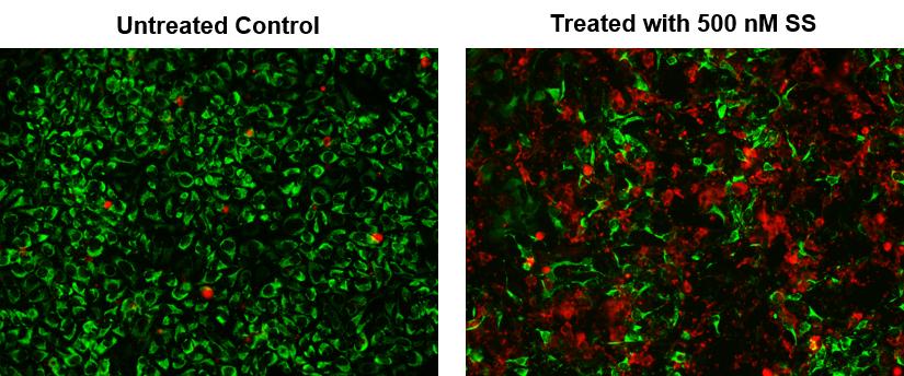
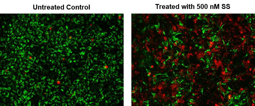
膜联蛋白V结合试剂盒旨在检测各种刺激诱导的细胞凋亡。它是基于细胞凋亡启动后膜磷脂酰丝氨酸(PS)从正常状态下的质膜内表面转移到细胞表面。细胞表面的PS可以通过用膜联蛋白V的荧光缀合物染色来检测,膜联蛋白V是一种对PS具有高亲和力的蛋白质。该检测可以通过流式细胞仪或荧光显微镜进行分析。当进行膜联蛋白V-FITC和PI染色时,该试剂盒可以区分凋亡和坏死。

关键特征

  • 快速:

    只需10-15分钟
  • 简单:

    单步
  • 可复制。

应用程序

  • 细胞凋亡
  • 细胞坏死

贮藏条件

将试剂储存在4°c下。保质期:6个月。

膜联蛋白V结合试验试剂盒(Annexin V Binding Assay)

重组Annexin V 蛋白 CAS 136107-94-3-AAT Bioquest荧光染料

上海金畔生物科技有限公司代理AAT Bioquest荧光染料全线产品,欢迎访问AAT Bioquest荧光染料官网了解更多信息。
重组Annexin V 蛋白 CAS 136107-94-3价格 2823
产品规格

100 ug

产品货号

重组Annexin V 蛋白 CAS 136107-94-3

产品参数
Ex (nm) Em (nm)
分子量 ~36000 溶剂 Water
存储条件 在零下15度以下保存, 避免光照
产品概述

产品基本信息

产品名称:膜联蛋白,重组

CAS:136107-94-3

储存条件:保存在冰箱-20℃干燥

来源:大肠杆菌

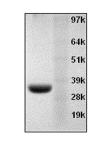
分子量:36 KD

纯度:通过SDS-PAGE> 95%

浓度:PBS中2 mg / mL(pH 7.4)

保质期:6个月


产品介绍

膜联蛋白V用作膜联蛋白V亲和力测定中的探针以检测凋亡细胞,其在细胞表面上表达磷脂酰丝氨酸,这是细胞凋亡以及其他形式的细胞死亡中发现的特征。 细胞凋亡是程序性细胞死亡的一种形式,其被身体用于从组织中去除不需要的,受损的或衰老的细胞。 通过白细胞如巨噬细胞或树突细胞的吞噬作用去除凋亡细胞。

膜联蛋白V已连续用于体外(培养管中的细胞)和体内(在实验室小鼠和患者中)检测凋亡细胞。 发生细胞凋亡的病理过程包括炎症,由心肌梗塞引起的心脏缺血损伤,血管中动脉粥样硬化斑块中存在的凋亡白细胞,供体患者中被免疫系统或肿瘤细胞排斥的移植器官。 化疗期间接触细胞生长抑制。

吞噬白细胞通过在细胞表面上暴露带负电荷的磷脂(磷脂酰丝氨酸)来识别凋亡细胞。在正常细胞中,阴性磷脂位于细胞膜的内侧,并且膜的外表面被磷脂占据,磷脂没有电荷。在细胞决定自杀后,带负电荷的磷脂通过称为scramblase的假想蛋白质转运到外细胞表面。吞噬白细胞表达受体,其可以检测带负电荷的磷脂。检测后,除去凋亡细胞。吞噬细胞迅速去除健康个体的凋亡细胞。然而,在病理过程中,凋亡细胞的去除可能被延迟或甚至不存在。可以用膜联蛋白V检测组织中的死亡细胞。

用荧光或放射性分子标记膜联蛋白V使得可以检测标记的膜联蛋白V与凋亡细胞的细胞表面的结合。在与磷脂表面结合后,膜联蛋白V组装成称为三聚体的簇。该三聚体由三个膜联蛋白V分子组成,它们通过非共价蛋白质 – 蛋白质相互作用彼此结合。膜联蛋白V三聚体的形成导致在磷脂膜上形成二维晶格。当用荧光或放射性探针标记时,膜上膜联蛋白V的聚集大大增加了膜联蛋白V的强度。


参考文献

Response of Mammalian cells to non-thermal intense narrowband pulsed electric fields
Authors: Sunao Katsuki, Yulan Li, Daiki Miyakawa, Ryo Yamada, Nobuaki Onishi, Soowon Lim
Journal: (2017): 1358–1361

Annexin V-生物素缀合物-AAT Bioquest荧光染料

上海金畔生物科技有限公司代理AAT Bioquest荧光染料全线产品,欢迎访问AAT Bioquest荧光染料官网了解更多信息。
Annexin V-生物素缀合物价格 2823
产品规格

100 tests

产品货号

Annexin V-生物素缀合物

产品参数
Ex (nm) Em (nm)
分子量 ~36000 溶剂 Water
存储条件 在零下15度以下保存, 避免光照
产品概述

Annexin V-生物素缀合物是美国AAT Bioquest生产的膜联蛋白,膜联蛋白是在钙存在下与磷脂膜结合的蛋白质家族。膜联蛋白V是研究细胞凋亡的有用工具。它被用作探针以检测在细胞表面上表达磷脂酰丝氨酸的细胞,这是细胞凋亡中发现的特征以及其他形式的细胞死亡。有多种参数可用于监测细胞活力。膜联蛋白V-染料缀合物广泛用于通过测量磷脂酰丝氨酸(PS)的易位来检测细胞凋亡。在细胞凋亡中,PS被转移到质膜的外部小叶。磷脂酰丝氨酸在细胞表面上的出现是细胞凋亡的初始/中间阶段的通用指示,并且可以在观察到形态变化之前检测到。Annexin V缀合物被广泛用于通过测量磷脂酰丝氨酸(PS)的转运来监测细胞凋亡。金畔生物是AAT Bioquest 的中国代理商,为您提供优质的Annexin V-生物素缀合物。

实验方案

操作步骤

1.用膜联蛋白V缀合物制备和孵育细胞:

1.1     制备膜联蛋白V缀合测定缓冲液:10mM HEPES,140mM NaCl和2.5mM CaCl2,pH 7.4。

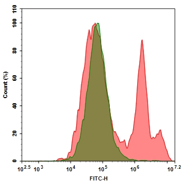
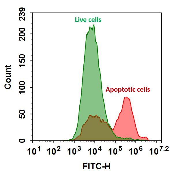
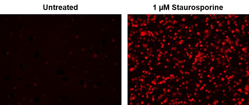
1.2     用测试化合物处理细胞一段时间(用星形孢菌素处理的Jurkat细胞4-6小时)以诱导细胞凋亡。

1.3     离心细胞,得到1-5×10 5个细胞/管。

1.4     将细胞重悬于200μLAnnexin V缀合测定缓冲液中(来自步骤1.1)。

1.5     在细胞中加入2μL膜联蛋白V缀合物。

可选:在细胞内加入死细胞染色剂如碘化丙锭,用于死细胞。

1.6     在室温下进行30至60分钟,避光。

1.7     在用流式细胞仪或荧光显微镜分析细胞之前,加入300μLAnnexin V缀合测定缓冲液(来自步骤1.1)以增加体积(参见下面的步骤1.8)。

1.8     使用流式细胞仪或荧光显微镜检测荧光强度(参见下面的步骤2或3)。


2.使用流式细胞仪分析:

通过使用具有适当滤波器的流式细胞仪来量化膜联蛋白V缀合物。

注意:对贴壁细胞的膜联蛋白V结合流式细胞术分析未经过常规测试,因为在细胞分离过程中可能会发生特定膜损伤。


3.使用荧光显微镜分析:

3.1    吸取步骤1.6中的细胞悬液,用膜联蛋白V结合测定缓冲液(步骤1.1)冲洗1-2次,然后用膜联蛋白V结合测定缓冲液(步骤1.1)重悬细胞。 将细胞添加到载玻片上并盖上盖玻片。

注意:对于贴壁细胞,建议直接在盖玻片上培养细胞。 与膜联蛋白V结合物孵育后(步骤1.6),用膜联蛋白V结合测定缓冲液(来自步骤1.1)冲洗1-2次,然后将膜联蛋白V结合测定缓冲液(来自步骤1.1)加回到盖玻片上。 将盖玻片倒在载玻片上并使细胞可视化。 与膜联蛋白V缀合物一起孵育后,也可以将细胞固定在2%甲醛中,并在显微镜下观察。

3.2     在荧光显微镜下用适当的滤光片分析膜联蛋白V缀合物的凋亡细胞。


参考文献

Response of Mammalian cells to non-thermal intense narrowband pulsed electric fields
Authors: Sunao Katsuki, Yulan Li, Daiki Miyakawa, Ryo Yamada, Nobuaki Onishi, Soowon Lim
Journal: (2017): 1358–1361


相关产品

产品名称 货号
Annexin V, TRITC标记 Cat#20031

Annexin V, FITC标记-AAT Bioquest荧光染料

上海金畔生物科技有限公司代理AAT Bioquest荧光染料全线产品,欢迎访问AAT Bioquest荧光染料官网了解更多信息。
Annexin V, FITC标记价格 2823
产品规格

100 tests

产品货号

Annexin V, FITC标记

产品参数
Ex (nm) 491 Em (nm) 516
分子量 ~36000 溶剂 Water
存储条件 在零下15度以下保存, 避免光照
产品概述

Annexin V, FITC标记是美国AAT Bioquest生产的膜联蛋白,膜联蛋白是在钙存在下与磷脂膜结合的蛋白质家族。膜联蛋白V是研究细胞凋亡的有用工具。它被用作探针以检测在细胞表面上表达磷脂酰丝氨酸的细胞,这是细胞凋亡中发现的特征以及其他形式的细胞死亡。有多种参数可用于监测细胞活力。膜联蛋白V-染料结合物广泛用于通过测量磷脂酰丝氨酸(PS)的易位来监测细胞凋亡。在细胞凋亡中,PS被转移到质膜的外部小叶。磷脂酰丝氨酸在细胞表面上的出现是细胞凋亡的初始/中间阶段的通用指示,并且可以在观察到形态变化之前检测到。该荧光FITC-膜联蛋白V缀合物广泛用于通过流式细胞仪或荧光显微镜监测细胞凋亡。对于基于膜联蛋白V的细胞凋亡检测的微孔板检测,我们建议您使用我们的Cell Meter 试剂盒。金畔生物是AAT Bioquest 的中国代理商,为您提供优质的Annexin V, FITC标记探针。

点击查看光谱

实验方案

操作步骤

1.用膜联蛋白V缀合物制备和孵育细胞:

1.1     制备膜联蛋白V缀合测定缓冲液:10mM HEPES,140mM NaCl和2.5mM CaCl2,pH 7.4。

1.2     用测试化合物处理细胞一段时间(用星形孢菌素处理的Jurkat细胞4-6小时)以诱导细胞凋亡。

1.3     离心细胞,得到1-5×10 5个细胞/管。

1.4     将细胞重悬于200μLAnnexin V缀合测定缓冲液中(来自步骤1.1)。

1.5     在细胞中加入2μL膜联蛋白V缀合物。

可选:在细胞内加入死细胞染色剂如碘化丙锭,用于死细胞。

1.6     在室温下进行30至60分钟,避光。

1.7     在用流式细胞仪或荧光显微镜分析细胞之前,加入300μLAnnexin V缀合测定缓冲液(来自步骤1.1)以增加体积(参见下面的步骤1.8)。

1.8     使用流式细胞仪或荧光显微镜检测荧光强度(参见下面的步骤2或3)。


2.使用流式细胞仪分析:

通过使用具有适当滤波器的流式细胞仪来量化膜联蛋白V缀合物。

注意:对贴壁细胞的膜联蛋白V结合流式细胞术分析未经过常规测试,因为在细胞分离过程中可能会发生特定膜损伤。


3.使用荧光显微镜分析:

3.1    吸取步骤1.6中的细胞悬液,用膜联蛋白V结合测定缓冲液(步骤1.1)冲洗1-2次,然后用膜联蛋白V结合测定缓冲液(步骤1.1)重悬细胞。 将细胞添加到载玻片上并盖上盖玻片。

注意:对于贴壁细胞,建议直接在盖玻片上培养细胞。 与膜联蛋白V结合物孵育后(步骤1.6),用膜联蛋白V结合测定缓冲液(来自步骤1.1)冲洗1-2次,然后将膜联蛋白V结合测定缓冲液(来自步骤1.1)加回到盖玻片上。 将盖玻片倒在载玻片上并使细胞可视化。 与膜联蛋白V缀合物一起孵育后,也可以将细胞固定在2%甲醛中,并在显微镜下观察。

3.2     在荧光显微镜下用适当的滤光片分析膜联蛋白V缀合物的凋亡细胞。


参考文献

Response of Mammalian cells to non-thermal intense narrowband pulsed electric fields
Authors: Sunao Katsuki, Yulan Li, Daiki Miyakawa, Ryo Yamada, Nobuaki Onishi, Soowon Lim
Journal: (2017): 1358–1361


相关产品

产品名称 货号
Annexin V, TRITC标记 Cat#20031

Annexin V, TRITC标记-AAT Bioquest荧光染料

上海金畔生物科技有限公司代理AAT Bioquest荧光染料全线产品,欢迎访问AAT Bioquest荧光染料官网了解更多信息。
Annexin V, TRITC标记价格 2823
产品规格

100 tests

产品货号

Annexin V, TRITC标记

产品参数
Ex (nm) 544 Em (nm) 570
分子量 ~36000 溶剂 Water
存储条件 在零下15度以下保存, 避免光照
产品概述

Annexin V, TRITC标记是美国AAT Bioquest生产的膜联蛋白,膜联蛋白是在钙存在下与磷脂膜结合的蛋白质家族。膜联蛋白V是研究细胞凋亡的有价值的工具。它用作检测在细胞表面表达磷脂酰丝氨酸的细胞的探针,这是细胞凋亡以及其他形式的细胞死亡中发现的特征。有多种参数可用于监测细胞活力。Annexin V-dye共轭物被广泛用于通过测量磷脂酰丝氨酸(PS)的转运来监测细胞凋亡。在细胞凋亡中,PS转移到质膜的外部小叶。磷脂酰丝氨酸在细胞表面的出现是细胞凋亡初始/中间阶段的普遍指标,可以在观察形态变化之前进行检测。这种荧光TRITC-Annexin V偶联物广泛用于通过流式细胞仪或荧光显微镜监测细胞凋亡。对于基于膜联蛋白V的细胞凋亡检测的微孔板检测,我们建议您使用Cell Meter™试剂盒。金畔生物是AAT Bioquest 的中国代理商,为您提供优质的Annexin V, TRITC标记探针。

点击查看光谱

实验方案

操作步骤

1.用膜联蛋白V缀合物制备和孵育细胞:

1.1     制备膜联蛋白V缀合测定缓冲液:10mM HEPES,140mM NaCl和2.5mM CaCl 2,pH 7.4。

1.2     用测试化合物处理细胞一段所需的时间(用星形孢菌素处理的Jurkat细胞4-6小时)以诱导细胞凋亡。

1.3     离心细胞,得到1-5×10 5个细胞/管。

1.4     将细胞重悬于200μLAnnexin V结合测定缓冲液中(来自步骤1.1)。

1.5     在细胞中加入2μL膜联蛋白V结合物。

可选:在细胞内加入死细胞染色剂如碘化丙锭,用于坏死细胞。

1.6     余ncubate在室温下进行30至60分钟,避光。

1.7     在用流式细胞仪或荧光显微镜分析细胞之前,加入300μLAnnexin V结合测定缓冲液(来自步骤1.1)以增加体积(参见下面的步骤1.8)。

1.8     使用流式细胞仪或荧光显微镜监测荧光强度(参见下面的步骤2或3)。


2.使用流式细胞仪分析:

通过使用具有适当过滤器的流式细胞仪来量化膜联蛋白V缀合物。

注意:粘附细胞上的膜联蛋白V 结合流式细胞术分析未经常检测,因为在细胞分离或收获期间可能发生特定的膜损伤。然而,Casiola-Rosen等人先前报道了利用膜联蛋白V对贴壁细胞类型进行流式细胞术的方法。和van Engelend等人(见参考文献1和2)。


3.使用荧光显微镜分析:

3.1     移液器从步骤1.6,将细胞悬浮液冲洗1-2次用甲nnexin V-结合测定缓冲液(来自步骤1.1) ,然后与A重悬细胞 nnexin V-结合测定缓冲液(来自步骤1.1) 。将细胞添加到盖有玻璃盖玻片的载玻片上。

注意:对于粘附细胞,建议直接在盖玻片上生长细胞。与膜联蛋白V缀合物(步骤1.6)孵育后,用Nnexin V结合测定缓冲液(来自步骤1.1)冲洗1-2次,并将A nnexin V结合测定缓冲液(来自步骤1.1)加回到盖玻片中。在玻璃载玻片上翻转盖玻片并观察细胞。在与膜联蛋白V缀合物孵育后,细胞也可以在2%甲醛中固定,并在显微镜下观察。

3.2     在荧光显微镜下用适当的滤光片分析膜联蛋白V缀合物的凋亡细胞。


参考文献

Response of Mammalian cells to non-thermal intense narrowband pulsed electric fields
Authors: Sunao Katsuki, Yulan Li, Daiki Miyakawa, Ryo Yamada, Nobuaki Onishi, Soowon Lim
Journal: (2017): 1358–1361


相关产品

产品名称 货号
Annexin V, FITC标记 Cat#20030

Annexin V-Cy3标记-AAT Bioquest荧光染料

上海金畔生物科技有限公司代理AAT Bioquest荧光染料全线产品,欢迎访问AAT Bioquest荧光染料官网了解更多信息。
Annexin V-Cy3标记价格 2823
产品规格

100 Tests

产品货号

Annexin V-Cy3标记

产品参数
Ex (nm) 555 Em (nm) 569
分子量 ~36000 溶剂 Water
存储条件 在零下15度以下保存, 避免光照
产品概述

Annexin V-Cy3标记是美国AAT Bioquest研发的用于检测细胞功能的探针,膜联蛋白是钙依赖性磷脂结合蛋白家族。 它们富含真核生物,属于参与信号转导的普遍存在的细胞质蛋白家族。 膜联蛋白V的优先结合配偶体是磷脂酰丝氨酸(PS),其通常保持在细胞膜的内叶(细胞质侧)。 在细胞凋亡中,PS被转移到质膜的外部小叶。 磷脂酰丝氨酸在细胞表面上的出现是细胞凋亡的初始/中间阶段的通用指示,并且可以在观察到形态变化之前检测到。 我们的膜联蛋白V结合物是一组监测细胞功能的工具,旨在通过测量磷脂酰丝氨酸的易位来监测细胞凋亡。

膜联蛋白V结合物在Ca2+存在下与凋亡细胞表面上的PS结合,它也可以通过坏死细胞膜或死细胞并与细胞内部的PS结合。 因此,我们建议将细胞不可渗透的核染色与膜联蛋白V结合物结合使用,以区分死细胞和凋亡细胞。金畔生物是AAT Bioquest的中国代理商,为您提供优质的Annexin V-Cy3检测试剂。 

点击查看光谱

实验方案

分析方案

概述

用测试化合物(200μL/样品)制备细胞

添加Annexin V结合物测定溶液

室温孵育30-60分钟

用流式细胞仪或荧光显微镜分析


1.用膜联蛋白V缀合物制备和孵育细胞:

1.1制备膜联蛋白V结合测定缓冲液:10mM HEPES,140mM NaCl和2.5mM CaCl2,pH 7.4。

1.2用测试化合物处理细胞一段所需的时间(用星形孢菌素处理的Jurkat细胞4-6小时)以诱导细胞凋亡。

1.3离心细胞,得到1-5×10⁵个细胞/管。

1.4将细胞重悬于200μL膜联蛋白V结合测定缓冲液中(来自步骤1.1)。

1.5在细胞中加入2μL膜联蛋白V结合物。

可选:在细胞内加入死细胞染色剂如碘化丙锭,用于坏死细胞。

1.6在室温下孵育30至60分钟,避光。

1.7在用流式细胞仪或荧光显微镜分析细胞之前,加入300μL膜联蛋白V结合测定缓冲液(来自步骤1.1)以增加体积(参见下面的步骤1.8)。

1.8使用流式细胞仪或荧光显微镜监测荧光强度(参见下面的步骤2或3)。


2.使用流式细胞仪分析:

通过使用具有适当过滤器的流式细胞仪来量化膜联蛋白V缀合物。

注意:粘附细胞上的膜联蛋白V结合流式细胞术分析未经常检测,因为在细胞分离或收获期间可能发生特定的膜损伤。 然而,Casiola-Rosen等人先前报道了利用膜联蛋白V对贴壁细胞类型进行流式细胞术的方法。 和van Engelend等人(见参考文献1和2)。


3.使用荧光显微镜分析:

3.1移取步骤1.6的细胞悬液,用膜联蛋白V结合测定缓冲液(来自步骤1.1)冲洗1-2次,然后用膜联蛋白V结合测定缓冲液(来自步骤1.1)重悬细胞。 将细胞添加到盖有玻璃盖玻片的载玻片上。

注意:对于粘附细胞,建议直接在盖玻片上生长细胞。 与膜联蛋白V缀合物孵育(步骤1.6)后,用膜联蛋白V结合测定缓冲液(来自步骤1.1)冲洗1-2次,并将膜联蛋白V结合测定缓冲液(来自步骤1.1)加回到盖玻片中。 在玻璃载玻片上翻转盖玻片并观察细胞。 在与膜联蛋白V缀合物孵育后,细胞也可以在2%甲醛中固定,并在显微镜下观察。

3.2在荧光显微镜下用适当的滤光片分析膜联蛋白V缀合物的凋亡细胞


数据分析

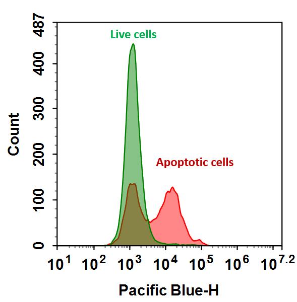
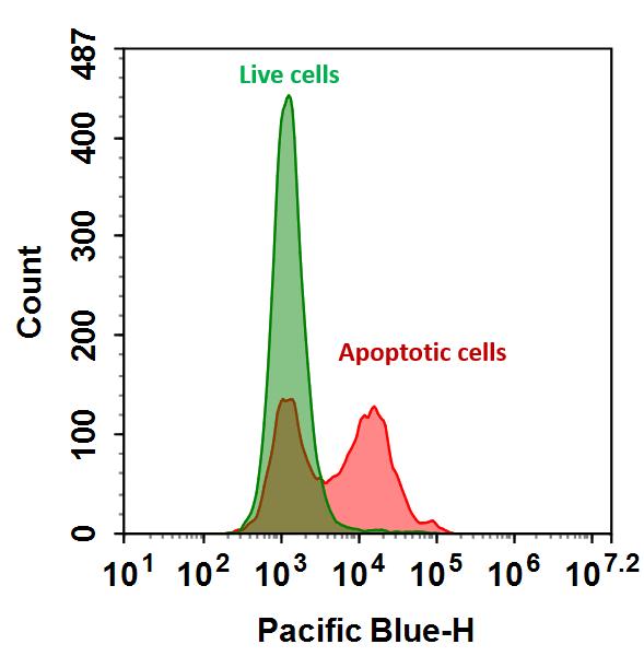
        下图是使用流式细胞仪检测膜联蛋白V-mFluor Violet 450与磷脂酰丝氨酸在Jurkat细胞凋亡中的结合活性的实例。 在活的非凋亡细胞中,膜联蛋白V-mFluor Violet 450检测非诱导细胞中的先天性细胞凋亡,其通常为所有细胞的2-6%。 在凋亡细胞中,膜联蛋白V-mFluor Violet 450与磷脂酰丝氨酸结合,磷脂酰丝氨酸位于细胞膜的外部小叶上,导致染色强度增加。

Annexin V-Cy3标记

图1.在Jurkat细胞中检测膜联蛋白VmFluor Violet 450和磷脂酰丝氨酸的结合活性。 将Jurkat细胞在37℃,5%CO2培养箱中不用(蓝色)或用1μM星形孢菌素(红色)处理5小时,然后用Annexin V-mFluor Violet 450染色30分钟。 使用FACSCalibur(Becton Dickinson,San Jose,CA)流式细胞仪,使用紫外激光在Ex / Em = 405 / 450nm下测量膜联蛋白V-mFluor Violet 450的荧光强度。


参考文献

Resources-Application Notes Phenotypic and Epigenetic Mechanism of Action Determinations of Histone Methylase and Demethylase Inhibitors using Digital Widefield Microscopy
Authors: Brad Larson, Peter Banks
Journal: Experimental Biology (2017)

Targeted Magnetic Intra-Lysosomal Hyperthermia produces lysosomal reactive oxygen species and causes Caspase-1 dependent cell death
Authors: Pascal Clerc, Pauline Jeanjean, Nicolas Halalli, Michel Gougeon, Bernard Pipy, Julian Carrey, Daniel Fourmy, Véronique Gigoux
Journal: Journal of Controlled Release (2017)

Annexin V-Cy5标记-AAT Bioquest荧光染料

上海金畔生物科技有限公司代理AAT Bioquest荧光染料全线产品,欢迎访问AAT Bioquest荧光染料官网了解更多信息。
Annexin V-Cy5标记价格 2823
产品规格

100 Tests

产品货号

Annexin V-Cy5标记

产品参数
Ex (nm) 656 Em (nm) 670
分子量 ~36000 溶剂 Water
存储条件 在零下15度以下保存, 避免光照
产品概述

Annexin V-Cy5标记是美国AAT Bioquest研发的用于检测细胞功能的探针,膜联蛋白是钙依赖性磷脂结合蛋白家族。 它们富含真核生物,属于参与信号转导的普遍存在的细胞质蛋白家族。 膜联蛋白V的优先结合配偶体是磷脂酰丝氨酸(PS),其通常保持在细胞膜的内叶(细胞质侧)。 在细胞凋亡中,PS被转移到质膜的外部小叶。 磷脂酰丝氨酸在细胞表面上的出现是细胞凋亡的初始/中间阶段的通用指示,并且可以在观察到形态变化之前检测到。 我们的膜联蛋白V结合物是一组监测细胞功能的工具,旨在通过测量磷脂酰丝氨酸的易位来监测细胞凋亡。

膜联蛋白V结合物在Ca2+存在下与凋亡细胞表面上的PS结合,它也可以通过坏死细胞膜或死细胞并与细胞内部的PS结合。 因此,我们建议将细胞不可渗透的核染色与膜联蛋白V结合物结合使用,以区分死细胞和凋亡细胞。金畔生物是AAT Bioquest的中国代理商,为您提供优质的Annexin V-Cy5检测试剂。 

点击查看光谱

实验方案

分析方案

概述

用测试化合物(200μL/样品)制备细胞

添加Annexin V结合物测定溶液

室温孵育30-60分钟

用流式细胞仪或荧光显微镜分析


1.用膜联蛋白V缀合物制备和孵育细胞:

1.1制备膜联蛋白V结合测定缓冲液:10mM HEPES,140mM NaCl和2.5mM CaCl2,pH 7.4。

1.2用测试化合物处理细胞一段所需的时间(用星形孢菌素处理的Jurkat细胞4-6小时)以诱导细胞凋亡。

1.3离心细胞,得到1-5×10⁵个细胞/管。

1.4将细胞重悬于200μL膜联蛋白V结合测定缓冲液中(来自步骤1.1)。

1.5在细胞中加入2μL膜联蛋白V结合物。

可选:在细胞内加入死细胞染色剂如碘化丙锭,用于坏死细胞。

1.6在室温下孵育30至60分钟,避光。

1.7在用流式细胞仪或荧光显微镜分析细胞之前,加入300μL膜联蛋白V结合测定缓冲液(来自步骤1.1)以增加体积(参见下面的步骤1.8)。

1.8使用流式细胞仪或荧光显微镜监测荧光强度(参见下面的步骤2或3)。


2.使用流式细胞仪分析:

通过使用具有适当过滤器的流式细胞仪来量化膜联蛋白V缀合物。

注意:粘附细胞上的膜联蛋白V结合流式细胞术分析未经常检测,因为在细胞分离或收获期间可能发生特定的膜损伤。 然而,Casiola-Rosen等人先前报道了利用膜联蛋白V对贴壁细胞类型进行流式细胞术的方法。 和van Engelend等人(见参考文献1和2)。


3.使用荧光显微镜分析:

3.1移取步骤1.6的细胞悬液,用膜联蛋白V结合测定缓冲液(来自步骤1.1)冲洗1-2次,然后用膜联蛋白V结合测定缓冲液(来自步骤1.1)重悬细胞。 将细胞添加到盖有玻璃盖玻片的载玻片上。

注意:对于粘附细胞,建议直接在盖玻片上生长细胞。 与膜联蛋白V缀合物孵育(步骤1.6)后,用膜联蛋白V结合测定缓冲液(来自步骤1.1)冲洗1-2次,并将膜联蛋白V结合测定缓冲液(来自步骤1.1)加回到盖玻片中。 在玻璃载玻片上翻转盖玻片并观察细胞。 在与膜联蛋白V缀合物孵育后,细胞也可以在2%甲醛中固定,并在显微镜下观察。

3.2在荧光显微镜下用适当的滤光片分析膜联蛋白V缀合物的凋亡细胞


数据分析

        下图是使用流式细胞仪检测膜联蛋白V-mFluor Violet 450与磷脂酰丝氨酸在Jurkat细胞凋亡中的结合活性的实例。 在活的非凋亡细胞中,膜联蛋白V-mFluor Violet 450检测非诱导细胞中的先天性细胞凋亡,其通常为所有细胞的2-6%。 在凋亡细胞中,膜联蛋白V-mFluor Violet 450与磷脂酰丝氨酸结合,磷脂酰丝氨酸位于细胞膜的外部小叶上,导致染色强度增加。

Annexin V-Cy5标记

图1.在Jurkat细胞中检测膜联蛋白VmFluor Violet 450和磷脂酰丝氨酸的结合活性。 将Jurkat细胞在37℃,5%CO2培养箱中不用(蓝色)或用1μM星形孢菌素(红色)处理5小时,然后用Annexin V-mFluor Violet 450染色30分钟。 使用FACSCalibur(Becton Dickinson,San Jose,CA)流式细胞仪,使用紫外激光在Ex / Em = 405 / 450nm下测量膜联蛋白V-mFluor Violet 450的荧光强度。


参考文献

Resources-Application Notes Phenotypic and Epigenetic Mechanism of Action Determinations of Histone Methylase and Demethylase Inhibitors using Digital Widefield Microscopy
Authors: Brad Larson, Peter Banks
Journal: Experimental Biology (2017)

Targeted Magnetic Intra-Lysosomal Hyperthermia produces lysosomal reactive oxygen species and causes Caspase-1 dependent cell death
Authors: Pascal Clerc, Pauline Jeanjean, Nicolas Halalli, Michel Gougeon, Bernard Pipy, Julian Carrey, Daniel Fourmy, Véronique Gigoux
Journal: Journal of Controlled Release (2017)

Annexin V-Cy5.5标记-AAT Bioquest荧光染料

上海金畔生物科技有限公司代理AAT Bioquest荧光染料全线产品,欢迎访问AAT Bioquest荧光染料官网了解更多信息。
Annexin V-Cy5.5标记价格 2823
产品规格

100 Tests

产品货号

Annexin V-Cy5.5标记

产品参数
Ex (nm) 684 Em (nm) 701
分子量 ~36000 溶剂 Water
存储条件 在零下15度以下保存, 避免光照
产品概述

Annexin V-Cy5.5标记是美国AAT Bioquest研发的用于检测细胞功能的探针,膜联蛋白是钙依赖性磷脂结合蛋白家族。 它们富含真核生物,属于参与信号转导的普遍存在的细胞质蛋白家族。 膜联蛋白V的优先结合配偶体是磷脂酰丝氨酸(PS),其通常保持在细胞膜的内叶(细胞质侧)。 在细胞凋亡中,PS被转移到质膜的外部小叶。 磷脂酰丝氨酸在细胞表面上的出现是细胞凋亡的初始/中间阶段的通用指示,并且可以在观察到形态变化之前检测到。 我们的膜联蛋白V结合物是一组监测细胞功能的工具,旨在通过测量磷脂酰丝氨酸的易位来监测细胞凋亡。

膜联蛋白V结合物在Ca2+存在下与凋亡细胞表面上的PS结合,它也可以通过坏死细胞膜或死细胞并与细胞内部的PS结合。 因此,我们建议将细胞不可渗透的核染色与膜联蛋白V结合物结合使用,以区分死细胞和凋亡细胞。金畔生物是AAT Bioquest的中国代理商,为您提供优质的Annexin V-Cy5.5检测试剂。 

点击查看光谱

实验方案

分析方案

概述

用测试化合物(200μL/样品)制备细胞

添加Annexin V结合物测定溶液

室温孵育30-60分钟

用流式细胞仪或荧光显微镜分析


1.用膜联蛋白V缀合物制备和孵育细胞:

1.1制备膜联蛋白V结合测定缓冲液:10mM HEPES,140mM NaCl和2.5mM CaCl2,pH 7.4。

1.2用测试化合物处理细胞一段所需的时间(用星形孢菌素处理的Jurkat细胞4-6小时)以诱导细胞凋亡。

1.3离心细胞,得到1-5×10⁵个细胞/管。

1.4将细胞重悬于200μL膜联蛋白V结合测定缓冲液中(来自步骤1.1)。

1.5在细胞中加入2μL膜联蛋白V结合物。

可选:在细胞内加入死细胞染色剂如碘化丙锭,用于坏死细胞。

1.6在室温下孵育30至60分钟,避光。

1.7在用流式细胞仪或荧光显微镜分析细胞之前,加入300μL膜联蛋白V结合测定缓冲液(来自步骤1.1)以增加体积(参见下面的步骤1.8)。

1.8使用流式细胞仪或荧光显微镜监测荧光强度(参见下面的步骤2或3)。


2.使用流式细胞仪分析:

通过使用具有适当过滤器的流式细胞仪来量化膜联蛋白V缀合物。

注意:粘附细胞上的膜联蛋白V结合流式细胞术分析未经常检测,因为在细胞分离或收获期间可能发生特定的膜损伤。 然而,Casiola-Rosen等人先前报道了利用膜联蛋白V对贴壁细胞类型进行流式细胞术的方法。 和van Engelend等人(见参考文献1和2)。


3.使用荧光显微镜分析:

3.1移取步骤1.6的细胞悬液,用膜联蛋白V结合测定缓冲液(来自步骤1.1)冲洗1-2次,然后用膜联蛋白V结合测定缓冲液(来自步骤1.1)重悬细胞。 将细胞添加到盖有玻璃盖玻片的载玻片上。

注意:对于粘附细胞,建议直接在盖玻片上生长细胞。 与膜联蛋白V缀合物孵育(步骤1.6)后,用膜联蛋白V结合测定缓冲液(来自步骤1.1)冲洗1-2次,并将膜联蛋白V结合测定缓冲液(来自步骤1.1)加回到盖玻片中。 在玻璃载玻片上翻转盖玻片并观察细胞。 在与膜联蛋白V缀合物孵育后,细胞也可以在2%甲醛中固定,并在显微镜下观察。

3.2在荧光显微镜下用适当的滤光片分析膜联蛋白V缀合物的凋亡细胞


数据分析

        下图是使用流式细胞仪检测膜联蛋白V-mFluor Violet 450与磷脂酰丝氨酸在Jurkat细胞凋亡中的结合活性的实例。 在活的非凋亡细胞中,膜联蛋白V-mFluor Violet 450检测非诱导细胞中的先天性细胞凋亡,其通常为所有细胞的2-6%。 在凋亡细胞中,膜联蛋白V-mFluor Violet 450与磷脂酰丝氨酸结合,磷脂酰丝氨酸位于细胞膜的外部小叶上,导致染色强度增加。

Annexin V-Cy5.5标记

图1.在Jurkat细胞中检测膜联蛋白VmFluor Violet 450和磷脂酰丝氨酸的结合活性。 将Jurkat细胞在37℃,5%CO2培养箱中不用(蓝色)或用1μM星形孢菌素(红色)处理5小时,然后用Annexin V-mFluor Violet 450染色30分钟。 使用FACSCalibur(Becton Dickinson,San Jose,CA)流式细胞仪,使用紫外激光在Ex / Em = 405 / 450nm下测量膜联蛋白V-mFluor Violet 450的荧光强度。


参考文献

Resources-Application Notes Phenotypic and Epigenetic Mechanism of Action Determinations of Histone Methylase and Demethylase Inhibitors using Digital Widefield Microscopy
Authors: Brad Larson, Peter Banks
Journal: Experimental Biology (2017)

Targeted Magnetic Intra-Lysosomal Hyperthermia produces lysosomal reactive oxygen species and causes Caspase-1 dependent cell death
Authors: Pascal Clerc, Pauline Jeanjean, Nicolas Halalli, Michel Gougeon, Bernard Pipy, Julian Carrey, Daniel Fourmy, Véronique Gigoux
Journal: Journal of Controlled Release (2017)

Annexin V-Cy7标记-AAT Bioquest荧光染料

上海金畔生物科技有限公司代理AAT Bioquest荧光染料全线产品,欢迎访问AAT Bioquest荧光染料官网了解更多信息。
Annexin V-Cy7标记价格 2823
产品规格

100 tests

产品货号

Annexin V-Cy7标记

产品参数
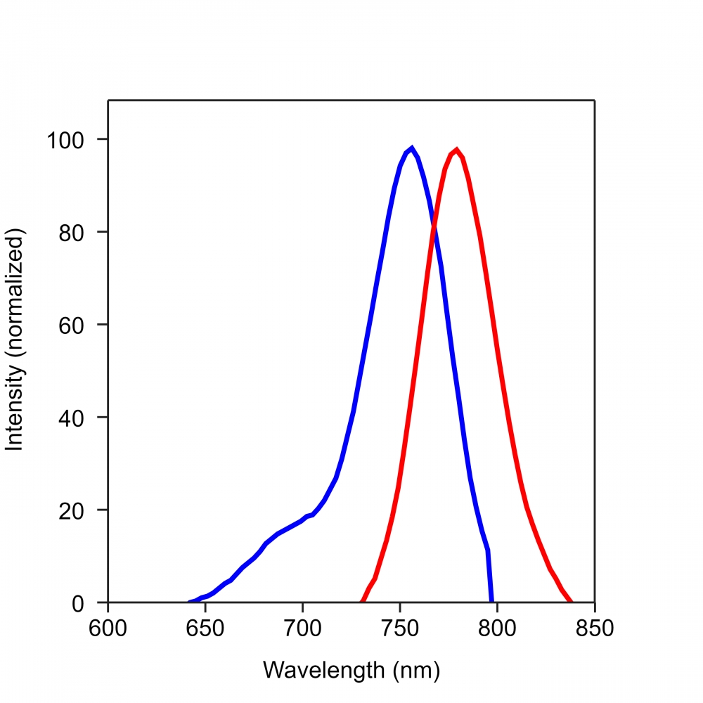
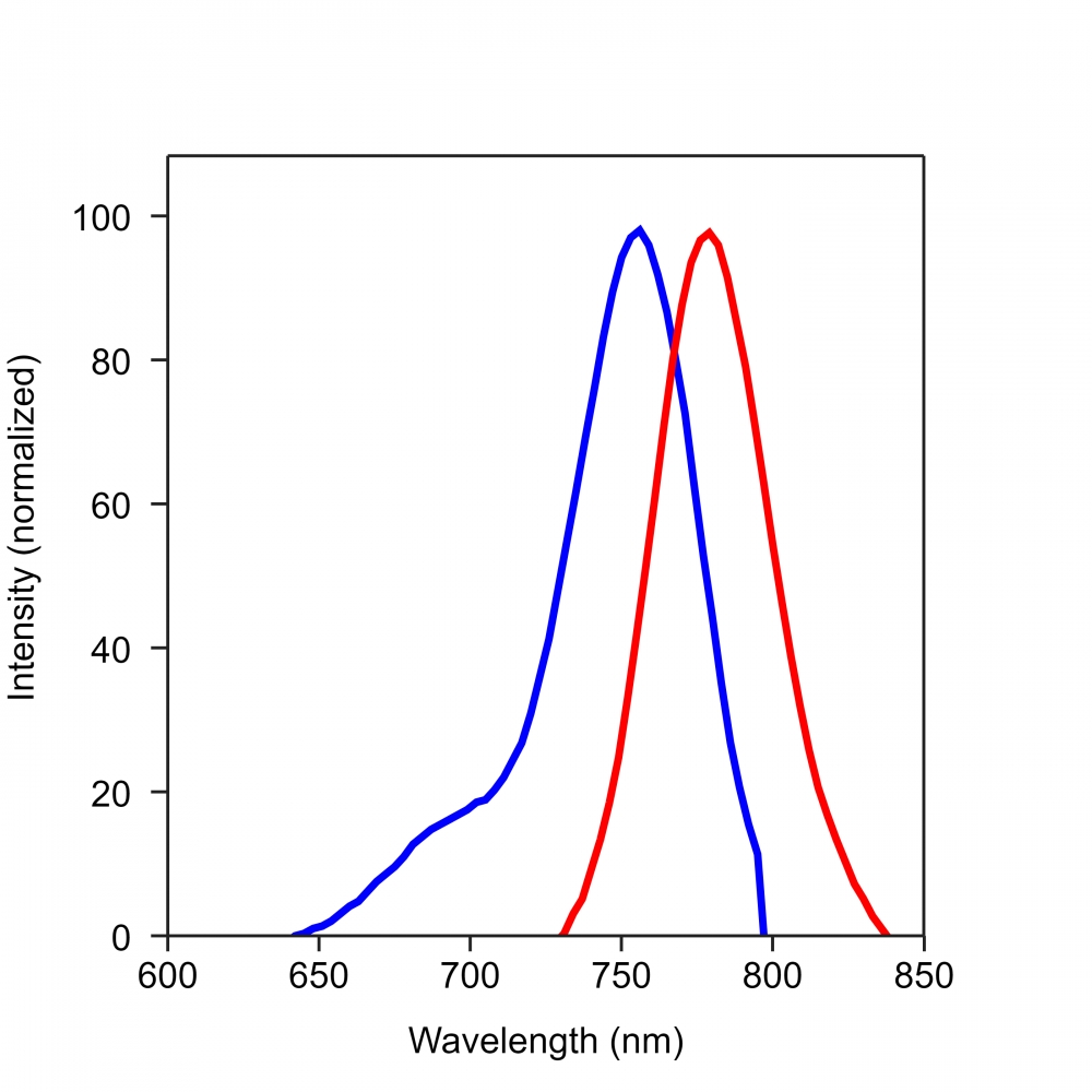
Ex (nm) 756 Em (nm) 779
分子量 ~36000 溶剂 Water
存储条件 在零下15度以下保存, 避免光照
产品概述

Annexin V-Cy7标记是美国AAT Bioquest研发的用于检测细胞功能的探针,膜联蛋白是钙依赖性磷脂结合蛋白家族。 它们富含真核生物,属于参与信号转导的普遍存在的细胞质蛋白家族。 膜联蛋白V的优先结合配偶体是磷脂酰丝氨酸(PS),其通常保持在细胞膜的内叶(细胞质侧)。 在细胞凋亡中,PS被转移到质膜的外部小叶。 磷脂酰丝氨酸在细胞表面上的出现是细胞凋亡的初始/中间阶段的通用指示,并且可以在观察到形态变化之前检测到。 我们的膜联蛋白V结合物是一组监测细胞功能的工具,旨在通过测量磷脂酰丝氨酸的易位来监测细胞凋亡。

膜联蛋白V结合物在Ca2+存在下与凋亡细胞表面上的PS结合,它也可以通过坏死细胞膜或死细胞并与细胞内部的PS结合。 因此,我们建议将细胞不可渗透的核染色与膜联蛋白V结合物结合使用,以区分死细胞和凋亡细胞。金畔生物是AAT Bioquest的中国代理商,为您提供优质的Annexin V-Cy7检测试剂。 

点击查看光谱

实验方案

分析方案

概述

用测试化合物(200μL/样品)制备细胞

添加Annexin V结合物测定溶液

室温孵育30-60分钟

用流式细胞仪或荧光显微镜分析


1.用膜联蛋白V缀合物制备和孵育细胞:

1.1制备膜联蛋白V结合测定缓冲液:10mM HEPES,140mM NaCl和2.5mM CaCl2,pH 7.4。

1.2用测试化合物处理细胞一段所需的时间(用星形孢菌素处理的Jurkat细胞4-6小时)以诱导细胞凋亡。

1.3离心细胞,得到1-5×10⁵个细胞/管。

1.4将细胞重悬于200μL膜联蛋白V结合测定缓冲液中(来自步骤1.1)。

1.5在细胞中加入2μL膜联蛋白V结合物。

可选:在细胞内加入死细胞染色剂如碘化丙锭,用于坏死细胞。

1.6在室温下孵育30至60分钟,避光。

1.7在用流式细胞仪或荧光显微镜分析细胞之前,加入300μL膜联蛋白V结合测定缓冲液(来自步骤1.1)以增加体积(参见下面的步骤1.8)。

1.8使用流式细胞仪或荧光显微镜监测荧光强度(参见下面的步骤2或3)。


2.使用流式细胞仪分析:

通过使用具有适当过滤器的流式细胞仪来量化膜联蛋白V缀合物。

注意:粘附细胞上的膜联蛋白V结合流式细胞术分析未经常检测,因为在细胞分离或收获期间可能发生特定的膜损伤。 然而,Casiola-Rosen等人先前报道了利用膜联蛋白V对贴壁细胞类型进行流式细胞术的方法。 和van Engelend等人(见参考文献1和2)。


3.使用荧光显微镜分析:

3.1移取步骤1.6的细胞悬液,用膜联蛋白V结合测定缓冲液(来自步骤1.1)冲洗1-2次,然后用膜联蛋白V结合测定缓冲液(来自步骤1.1)重悬细胞。 将细胞添加到盖有玻璃盖玻片的载玻片上。

注意:对于粘附细胞,建议直接在盖玻片上生长细胞。 与膜联蛋白V缀合物孵育(步骤1.6)后,用膜联蛋白V结合测定缓冲液(来自步骤1.1)冲洗1-2次,并将膜联蛋白V结合测定缓冲液(来自步骤1.1)加回到盖玻片中。 在玻璃载玻片上翻转盖玻片并观察细胞。 在与膜联蛋白V缀合物孵育后,细胞也可以在2%甲醛中固定,并在显微镜下观察。

3.2在荧光显微镜下用适当的滤光片分析膜联蛋白V缀合物的凋亡细胞


数据分析

        下图是使用流式细胞仪检测膜联蛋白V-mFluor Violet 450与磷脂酰丝氨酸在Jurkat细胞凋亡中的结合活性的实例。 在活的非凋亡细胞中,膜联蛋白V-mFluor Violet 450检测非诱导细胞中的先天性细胞凋亡,其通常为所有细胞的2-6%。 在凋亡细胞中,膜联蛋白V-mFluor Violet 450与磷脂酰丝氨酸结合,磷脂酰丝氨酸位于细胞膜的外部小叶上,导致染色强度增加。

Annexin V-Cy7标记

图1.在Jurkat细胞中检测膜联蛋白VmFluor Violet 450和磷脂酰丝氨酸的结合活性。 将Jurkat细胞在37℃,5%CO2培养箱中不用(蓝色)或用1μM星形孢菌素(红色)处理5小时,然后用Annexin V-mFluor Violet 450染色30分钟。 使用FACSCalibur(Becton Dickinson,San Jose,CA)流式细胞仪,使用紫外激光在Ex / Em = 405 / 450nm下测量膜联蛋白V-mFluor Violet 450的荧光强度。


参考文献

Resources-Application Notes Phenotypic and Epigenetic Mechanism of Action Determinations of Histone Methylase and Demethylase Inhibitors using Digital Widefield Microscopy
Authors: Brad Larson, Peter Banks
Journal: Experimental Biology (2017)

Targeted Magnetic Intra-Lysosomal Hyperthermia produces lysosomal reactive oxygen species and causes Caspase-1 dependent cell death
Authors: Pascal Clerc, Pauline Jeanjean, Nicolas Halalli, Michel Gougeon, Bernard Pipy, Julian Carrey, Daniel Fourmy, Véronique Gigoux
Journal: Journal of Controlled Release (2017)

Annexin V-iFluor 633标记-AAT Bioquest荧光染料

上海金畔生物科技有限公司代理AAT Bioquest荧光染料全线产品,欢迎访问AAT Bioquest荧光染料官网了解更多信息。
Annexin V-iFluor 633标记价格 2823
产品规格

100 tests

产品货号

Annexin V-iFluor 633标记

产品参数
Ex (nm) 640 Em (nm) 654
分子量 ~36000 溶剂 Water
存储条件 在零下15度以下保存, 避免光照
产品概述

Annexin V-iFluor 633标记是美国AAT Bioquest研发的用于检测细胞功能的探针,膜联蛋白是钙依赖性磷脂结合蛋白家族。 它们富含真核生物,属于参与信号转导的普遍存在的细胞质蛋白家族。 膜联蛋白V的优先结合配偶体是磷脂酰丝氨酸(PS),其通常保持在细胞膜的内叶(细胞质侧)。 在细胞凋亡中,PS被转移到质膜的外部小叶。 磷脂酰丝氨酸在细胞表面上的出现是细胞凋亡的初始/中间阶段的通用指示,并且可以在观察到形态变化之前检测到。 我们的膜联蛋白V结合物是一组监测细胞功能的工具,旨在通过测量磷脂酰丝氨酸的易位来监测细胞凋亡。

膜联蛋白V结合物在Ca2+存在下与凋亡细胞表面上的PS结合,它也可以通过坏死细胞膜或死细胞并与细胞内部的PS结合。 因此,我们建议将细胞不可渗透的核染色与膜联蛋白V结合物结合使用,以区分死细胞和凋亡细胞。金畔生物是AAT Bioquest的中国代理商,为您提供优质的Annexin V-iFluor 633检测试剂。 

点击查看光谱

实验方案

分析方案

概述

用测试化合物(200μL/样品)制备细胞

添加Annexin V结合物测定溶液

室温孵育30-60分钟

用流式细胞仪或荧光显微镜分析


1.用膜联蛋白V缀合物制备和孵育细胞:

1.1制备膜联蛋白V结合测定缓冲液:10mM HEPES,140mM NaCl和2.5mM CaCl2,pH 7.4。

1.2用测试化合物处理细胞一段所需的时间(用星形孢菌素处理的Jurkat细胞4-6小时)以诱导细胞凋亡。

1.3离心细胞,得到1-5×10⁵个细胞/管。

1.4将细胞重悬于200μL膜联蛋白V结合测定缓冲液中(来自步骤1.1)。

1.5在细胞中加入2μL膜联蛋白V结合物。

可选:在细胞内加入死细胞染色剂如碘化丙锭,用于坏死细胞。

1.6在室温下孵育30至60分钟,避光。

1.7在用流式细胞仪或荧光显微镜分析细胞之前,加入300μL膜联蛋白V结合测定缓冲液(来自步骤1.1)以增加体积(参见下面的步骤1.8)。

1.8使用流式细胞仪或荧光显微镜监测荧光强度(参见下面的步骤2或3)。


2.使用流式细胞仪分析:

通过使用具有适当过滤器的流式细胞仪来量化膜联蛋白V缀合物。

注意:粘附细胞上的膜联蛋白V结合流式细胞术分析未经常检测,因为在细胞分离或收获期间可能发生特定的膜损伤。 然而,Casiola-Rosen等人先前报道了利用膜联蛋白V对贴壁细胞类型进行流式细胞术的方法。 和van Engelend等人(见参考文献1和2)。


3.使用荧光显微镜分析:

3.1移取步骤1.6的细胞悬液,用膜联蛋白V结合测定缓冲液(来自步骤1.1)冲洗1-2次,然后用膜联蛋白V结合测定缓冲液(来自步骤1.1)重悬细胞。 将细胞添加到盖有玻璃盖玻片的载玻片上。

注意:对于粘附细胞,建议直接在盖玻片上生长细胞。 与膜联蛋白V缀合物孵育(步骤1.6)后,用膜联蛋白V结合测定缓冲液(来自步骤1.1)冲洗1-2次,并将膜联蛋白V结合测定缓冲液(来自步骤1.1)加回到盖玻片中。 在玻璃载玻片上翻转盖玻片并观察细胞。 在与膜联蛋白V缀合物孵育后,细胞也可以在2%甲醛中固定,并在显微镜下观察。

3.2在荧光显微镜下用适当的滤光片分析膜联蛋白V缀合物的凋亡细胞


数据分析

        下图是使用流式细胞仪检测膜联蛋白V-mFluor Violet 450与磷脂酰丝氨酸在Jurkat细胞凋亡中的结合活性的实例。 在活的非凋亡细胞中,膜联蛋白V-mFluor Violet 450检测非诱导细胞中的先天性细胞凋亡,其通常为所有细胞的2-6%。 在凋亡细胞中,膜联蛋白V-mFluor Violet 450与磷脂酰丝氨酸结合,磷脂酰丝氨酸位于细胞膜的外部小叶上,导致染色强度增加。

Annexin V-iFluor 633标记

图1.在Jurkat细胞中检测膜联蛋白VmFluor Violet 450和磷脂酰丝氨酸的结合活性。 将Jurkat细胞在37℃,5%CO2培养箱中不用(蓝色)或用1μM星形孢菌素(红色)处理5小时,然后用Annexin V-mFluor Violet 450染色30分钟。 使用FACSCalibur(Becton Dickinson,San Jose,CA)流式细胞仪,使用紫外激光在Ex / Em = 405 / 450nm下测量膜联蛋白V-mFluor Violet 450的荧光强度。


参考文献

Resources-Application Notes Phenotypic and Epigenetic Mechanism of Action Determinations of Histone Methylase and Demethylase Inhibitors using Digital Widefield Microscopy
Authors: Brad Larson, Peter Banks
Journal: Experimental Biology (2017)

Targeted Magnetic Intra-Lysosomal Hyperthermia produces lysosomal reactive oxygen species and causes Caspase-1 dependent cell death
Authors: Pascal Clerc, Pauline Jeanjean, Nicolas Halalli, Michel Gougeon, Bernard Pipy, Julian Carrey, Daniel Fourmy, Véronique Gigoux
Journal: Journal of Controlled Release (2017)

Annexin V-iFluor 350标记-AAT Bioquest荧光染料

上海金畔生物科技有限公司代理AAT Bioquest荧光染料全线产品,欢迎访问AAT Bioquest荧光染料官网了解更多信息。
Annexin V-iFluor 350标记价格 2823
产品规格

100 tests

产品货号

Annexin V-iFluor 350标记

产品参数
Ex (nm) 345 Em (nm) 450
分子量 ~36000 溶剂 Water
存储条件 在零下15度以下保存, 避免光照
产品概述

Annexin V-iFluor 350标记是美国AAT Bioquest研发的用于检测细胞功能的探针,膜联蛋白是钙依赖性磷脂结合蛋白家族。 它们富含真核生物,属于参与信号转导的普遍存在的细胞质蛋白家族。 膜联蛋白V的优先结合配偶体是磷脂酰丝氨酸(PS),其通常保持在细胞膜的内叶(细胞质侧)。 在细胞凋亡中,PS被转移到质膜的外部小叶。 磷脂酰丝氨酸在细胞表面上的出现是细胞凋亡的初始/中间阶段的通用指示,并且可以在观察到形态变化之前检测到。 我们的膜联蛋白V结合物是一组监测细胞功能的工具,旨在通过测量磷脂酰丝氨酸的易位来监测细胞凋亡。

膜联蛋白V结合物在Ca2+存在下与凋亡细胞表面上的PS结合,它也可以通过坏死细胞膜或死细胞并与细胞内部的PS结合。 因此,我们建议将细胞不可渗透的核染色与膜联蛋白V结合物结合使用,以区分死细胞和凋亡细胞。金畔生物是AAT Bioquest的中国代理商,为您提供优质的Annexin V-iFluor 350检测试剂。 


适用仪器


流式细胞仪  
Ex: 355 nm
Em: 450/50  nm 
通道: Pacific Blue


荧光显微镜

推荐孔板: 黑色透明底板
通道: Pacific Blue 通道
实验方案

分析方案

概述

用测试化合物(200μL/样品)制备细胞

添加Annexin V结合物测定溶液

室温孵育30-60分钟

用流式细胞仪或荧光显微镜分析


1.用膜联蛋白V缀合物制备和孵育细胞:

1.1制备膜联蛋白V结合测定缓冲液:10mM HEPES,140mM NaCl和2.5mM CaCl2,pH 7.4。

1.2用测试化合物处理细胞一段所需的时间(用星形孢菌素处理的Jurkat细胞4-6小时)以诱导细胞凋亡。

1.3离心细胞,得到1-5×10⁵个细胞/管。

1.4将细胞重悬于200μL膜联蛋白V结合测定缓冲液中(来自步骤1.1)。

1.5在细胞中加入2μL膜联蛋白V结合物。

可选:在细胞内加入死细胞染色剂如碘化丙锭,用于坏死细胞。

1.6在室温下孵育30至60分钟,避光。

1.7在用流式细胞仪或荧光显微镜分析细胞之前,加入300μL膜联蛋白V结合测定缓冲液(来自步骤1.1)以增加体积(参见下面的步骤1.8)。

1.8使用流式细胞仪或荧光显微镜监测荧光强度(参见下面的步骤2或3)。


2.使用流式细胞仪分析:

通过使用具有适当过滤器的流式细胞仪来量化膜联蛋白V缀合物。

注意:粘附细胞上的膜联蛋白V结合流式细胞术分析未经常检测,因为在细胞分离或收获期间可能发生特定的膜损伤。 然而,Casiola-Rosen等人先前报道了利用膜联蛋白V对贴壁细胞类型进行流式细胞术的方法。 和van Engelend等人(见参考文献1和2)。


3.使用荧光显微镜分析:

3.1移取步骤1.6的细胞悬液,用膜联蛋白V结合测定缓冲液(来自步骤1.1)冲洗1-2次,然后用膜联蛋白V结合测定缓冲液(来自步骤1.1)重悬细胞。 将细胞添加到盖有玻璃盖玻片的载玻片上。

注意:对于粘附细胞,建议直接在盖玻片上生长细胞。 与膜联蛋白V缀合物孵育(步骤1.6)后,用膜联蛋白V结合测定缓冲液(来自步骤1.1)冲洗1-2次,并将膜联蛋白V结合测定缓冲液(来自步骤1.1)加回到盖玻片中。 在玻璃载玻片上翻转盖玻片并观察细胞。 在与膜联蛋白V缀合物孵育后,细胞也可以在2%甲醛中固定,并在显微镜下观察。

3.2在荧光显微镜下用适当的滤光片分析膜联蛋白V缀合物的凋亡细胞


数据分析

        下图是使用流式细胞仪检测膜联蛋白V-mFluor Violet 450与磷脂酰丝氨酸在Jurkat细胞凋亡中的结合活性的实例。 在活的非凋亡细胞中,膜联蛋白V-mFluor Violet 450检测非诱导细胞中的先天性细胞凋亡,其通常为所有细胞的2-6%。 在凋亡细胞中,膜联蛋白V-mFluor Violet 450与磷脂酰丝氨酸结合,磷脂酰丝氨酸位于细胞膜的外部小叶上,导致染色强度增加。

Annexin V-iFluor 350标记

图1.在Jurkat细胞中检测膜联蛋白VmFluor Violet 450和磷脂酰丝氨酸的结合活性。 将Jurkat细胞在37℃,5%CO2培养箱中不用(蓝色)或用1μM星形孢菌素(红色)处理5小时,然后用Annexin V-mFluor Violet 450染色30分钟。 使用FACSCalibur(Becton Dickinson,San Jose,CA)流式细胞仪,使用紫外激光在Ex / Em = 405 / 450nm下测量膜联蛋白V-mFluor Violet 450的荧光强度。


参考文献

Resources-Application Notes Phenotypic and Epigenetic Mechanism of Action Determinations of Histone Methylase and Demethylase Inhibitors using Digital Widefield Microscopy
Authors: Brad Larson, Peter Banks
Journal: Experimental Biology (2017)

Targeted Magnetic Intra-Lysosomal Hyperthermia produces lysosomal reactive oxygen species and causes Caspase-1 dependent cell death
Authors: Pascal Clerc, Pauline Jeanjean, Nicolas Halalli, Michel Gougeon, Bernard Pipy, Julian Carrey, Daniel Fourmy, Véronique Gigoux
Journal: Journal of Controlled Release (2017)

Annexin V-iFluor 488标记-AAT Bioquest荧光染料

上海金畔生物科技有限公司代理AAT Bioquest荧光染料全线产品,欢迎访问AAT Bioquest荧光染料官网了解更多信息。
Annexin V-iFluor 488标记价格 2823
产品规格

100 tests

产品货号

Annexin V-iFluor 488标记

产品参数
Ex (nm) 491 Em (nm) 516
分子量 ~36000 溶剂 Water
存储条件 在零下15度以下保存, 避免光照
产品概述

Annexin V-iFluor 488标记是美国AAT Bioquest研发的用于检测细胞功能的探针,膜联蛋白是钙依赖性磷脂结合蛋白家族。 它们富含真核生物,属于参与信号转导的普遍存在的细胞质蛋白家族。 膜联蛋白V的优先结合配偶体是磷脂酰丝氨酸(PS),其通常保持在细胞膜的内叶(细胞质侧)。 在细胞凋亡中,PS被转移到质膜的外部小叶。 磷脂酰丝氨酸在细胞表面上的出现是细胞凋亡的初始/中间阶段的通用指示,并且可以在观察到形态变化之前检测到。 我们的膜联蛋白V结合物是一组监测细胞功能的工具,旨在通过测量磷脂酰丝氨酸的易位来监测细胞凋亡。

膜联蛋白V结合物在Ca2+存在下与凋亡细胞表面上的PS结合,它也可以通过坏死细胞膜或死细胞并与细胞内部的PS结合。 因此,我们建议将细胞不可渗透的核染色与膜联蛋白V结合物结合使用,以区分死细胞和凋亡细胞。金畔生物是AAT Bioquest的中国代理商,为您提供优质的Annexin V-iFluor 488检测试剂。

点击查看光谱 

实验方案

分析方案

概述

用测试化合物(200μL/样品)制备细胞

添加Annexin V结合物测定溶液

室温孵育30-60分钟

用流式细胞仪或荧光显微镜分析


1.用膜联蛋白V缀合物制备和孵育细胞:

1.1制备膜联蛋白V结合测定缓冲液:10mM HEPES,140mM NaCl和2.5mM CaCl2,pH 7.4。

1.2用测试化合物处理细胞一段所需的时间(用星形孢菌素处理的Jurkat细胞4-6小时)以诱导细胞凋亡。

1.3离心细胞,得到1-5×10⁵个细胞/管。

1.4将细胞重悬于200μL膜联蛋白V结合测定缓冲液中(来自步骤1.1)。

1.5在细胞中加入2μL膜联蛋白V结合物。

可选:在细胞内加入死细胞染色剂如碘化丙锭,用于坏死细胞。

1.6在室温下孵育30至60分钟,避光。

1.7在用流式细胞仪或荧光显微镜分析细胞之前,加入300μL膜联蛋白V结合测定缓冲液(来自步骤1.1)以增加体积(参见下面的步骤1.8)。

1.8使用流式细胞仪或荧光显微镜监测荧光强度(参见下面的步骤2或3)。


2.使用流式细胞仪分析:

通过使用具有适当过滤器的流式细胞仪来量化膜联蛋白V缀合物。

注意:粘附细胞上的膜联蛋白V结合流式细胞术分析未经常检测,因为在细胞分离或收获期间可能发生特定的膜损伤。 然而,Casiola-Rosen等人先前报道了利用膜联蛋白V对贴壁细胞类型进行流式细胞术的方法。 和van Engelend等人(见参考文献1和2)。


3.使用荧光显微镜分析:

3.1移取步骤1.6的细胞悬液,用膜联蛋白V结合测定缓冲液(来自步骤1.1)冲洗1-2次,然后用膜联蛋白V结合测定缓冲液(来自步骤1.1)重悬细胞。 将细胞添加到盖有玻璃盖玻片的载玻片上。

注意:对于粘附细胞,建议直接在盖玻片上生长细胞。 与膜联蛋白V缀合物孵育(步骤1.6)后,用膜联蛋白V结合测定缓冲液(来自步骤1.1)冲洗1-2次,并将膜联蛋白V结合测定缓冲液(来自步骤1.1)加回到盖玻片中。 在玻璃载玻片上翻转盖玻片并观察细胞。 在与膜联蛋白V缀合物孵育后,细胞也可以在2%甲醛中固定,并在显微镜下观察。

3.2在荧光显微镜下用适当的滤光片分析膜联蛋白V缀合物的凋亡细胞


数据分析

        下图是使用流式细胞仪检测膜联蛋白V-mFluor Violet 450与磷脂酰丝氨酸在Jurkat细胞凋亡中的结合活性的实例。 在活的非凋亡细胞中,膜联蛋白V-mFluor Violet 450检测非诱导细胞中的先天性细胞凋亡,其通常为所有细胞的2-6%。 在凋亡细胞中,膜联蛋白V-mFluor Violet 450与磷脂酰丝氨酸结合,磷脂酰丝氨酸位于细胞膜的外部小叶上,导致染色强度增加。

Annexin V-iFluor 488标记

图1.在Jurkat细胞中检测膜联蛋白VmFluor Violet 450和磷脂酰丝氨酸的结合活性。 将Jurkat细胞在37℃,5%CO2培养箱中不用(蓝色)或用1μM星形孢菌素(红色)处理5小时,然后用Annexin V-mFluor Violet 450染色30分钟。 使用FACSCalibur(Becton Dickinson,San Jose,CA)流式细胞仪,使用紫外激光在Ex / Em = 405 / 450nm下测量膜联蛋白V-mFluor Violet 450的荧光强度。


参考文献

Resources-Application Notes Phenotypic and Epigenetic Mechanism of Action Determinations of Histone Methylase and Demethylase Inhibitors using Digital Widefield Microscopy
Authors: Brad Larson, Peter Banks
Journal: Experimental Biology (2017)

Targeted Magnetic Intra-Lysosomal Hyperthermia produces lysosomal reactive oxygen species and causes Caspase-1 dependent cell death
Authors: Pascal Clerc, Pauline Jeanjean, Nicolas Halalli, Michel Gougeon, Bernard Pipy, Julian Carrey, Daniel Fourmy, Véronique Gigoux
Journal: Journal of Controlled Release (2017)

Annexin V-iFluor 555标记-AAT Bioquest荧光染料

上海金畔生物科技有限公司代理AAT Bioquest荧光染料全线产品,欢迎访问AAT Bioquest荧光染料官网了解更多信息。
Annexin V-iFluor 555标记价格 2823
产品规格

100 tests

产品货号

Annexin V-iFluor 555标记

产品参数
Ex (nm) 557 Em (nm) 570
分子量 ~36000 溶剂 Water
存储条件 在零下15度以下保存, 避免光照
产品概述

Annexin V-iFluor 555标记是美国AAT Bioquest研发的用于检测细胞功能的探针,膜联蛋白是钙依赖性磷脂结合蛋白家族。 它们富含真核生物,属于参与信号转导的普遍存在的细胞质蛋白家族。 膜联蛋白V的优先结合配偶体是磷脂酰丝氨酸(PS),其通常保持在细胞膜的内叶(细胞质侧)。 在细胞凋亡中,PS被转移到质膜的外部小叶。 磷脂酰丝氨酸在细胞表面上的出现是细胞凋亡的初始/中间阶段的通用指示,并且可以在观察到形态变化之前检测到。 我们的膜联蛋白V结合物是一组监测细胞功能的工具,旨在通过测量磷脂酰丝氨酸的易位来监测细胞凋亡。

膜联蛋白V结合物在Ca2+存在下与凋亡细胞表面上的PS结合,它也可以通过坏死细胞膜或死细胞并与细胞内部的PS结合。 因此,我们建议将细胞不可渗透的核染色与膜联蛋白V结合物结合使用,以区分死细胞和凋亡细胞。金畔生物是AAT Bioquest的中国代理商,为您提供优质的Annexin V-iFluor 555检测试剂。 

点击查看光谱

实验方案

分析方案

概述

用测试化合物(200μL/样品)制备细胞

添加Annexin V结合物测定溶液

室温孵育30-60分钟

用流式细胞仪或荧光显微镜分析


1.用膜联蛋白V缀合物制备和孵育细胞:

1.1制备膜联蛋白V结合测定缓冲液:10mM HEPES,140mM NaCl和2.5mM CaCl2,pH 7.4。

1.2用测试化合物处理细胞一段所需的时间(用星形孢菌素处理的Jurkat细胞4-6小时)以诱导细胞凋亡。

1.3离心细胞,得到1-5×10⁵个细胞/管。

1.4将细胞重悬于200μL膜联蛋白V结合测定缓冲液中(来自步骤1.1)。

1.5在细胞中加入2μL膜联蛋白V结合物。

可选:在细胞内加入死细胞染色剂如碘化丙锭,用于坏死细胞。

1.6在室温下孵育30至60分钟,避光。

1.7在用流式细胞仪或荧光显微镜分析细胞之前,加入300μL膜联蛋白V结合测定缓冲液(来自步骤1.1)以增加体积(参见下面的步骤1.8)。

1.8使用流式细胞仪或荧光显微镜监测荧光强度(参见下面的步骤2或3)。


2.使用流式细胞仪分析:

通过使用具有适当过滤器的流式细胞仪来量化膜联蛋白V缀合物。

注意:粘附细胞上的膜联蛋白V结合流式细胞术分析未经常检测,因为在细胞分离或收获期间可能发生特定的膜损伤。 然而,Casiola-Rosen等人先前报道了利用膜联蛋白V对贴壁细胞类型进行流式细胞术的方法。 和van Engelend等人(见参考文献1和2)。


3.使用荧光显微镜分析:

3.1移取步骤1.6的细胞悬液,用膜联蛋白V结合测定缓冲液(来自步骤1.1)冲洗1-2次,然后用膜联蛋白V结合测定缓冲液(来自步骤1.1)重悬细胞。 将细胞添加到盖有玻璃盖玻片的载玻片上。

注意:对于粘附细胞,建议直接在盖玻片上生长细胞。 与膜联蛋白V缀合物孵育(步骤1.6)后,用膜联蛋白V结合测定缓冲液(来自步骤1.1)冲洗1-2次,并将膜联蛋白V结合测定缓冲液(来自步骤1.1)加回到盖玻片中。 在玻璃载玻片上翻转盖玻片并观察细胞。 在与膜联蛋白V缀合物孵育后,细胞也可以在2%甲醛中固定,并在显微镜下观察。

3.2在荧光显微镜下用适当的滤光片分析膜联蛋白V缀合物的凋亡细胞


数据分析

        下图是使用流式细胞仪检测膜联蛋白V-mFluor Violet 450与磷脂酰丝氨酸在Jurkat细胞凋亡中的结合活性的实例。 在活的非凋亡细胞中,膜联蛋白V-mFluor Violet 450检测非诱导细胞中的先天性细胞凋亡,其通常为所有细胞的2-6%。 在凋亡细胞中,膜联蛋白V-mFluor Violet 450与磷脂酰丝氨酸结合,磷脂酰丝氨酸位于细胞膜的外部小叶上,导致染色强度增加。

Annexin V-iFluor 555标记

图1.在Jurkat细胞中检测膜联蛋白VmFluor Violet 450和磷脂酰丝氨酸的结合活性。 将Jurkat细胞在37℃,5%CO2培养箱中不用(蓝色)或用1μM星形孢菌素(红色)处理5小时,然后用Annexin V-mFluor Violet 450染色30分钟。 使用FACSCalibur(Becton Dickinson,San Jose,CA)流式细胞仪,使用紫外激光在Ex / Em = 405 / 450nm下测量膜联蛋白V-mFluor Violet 450的荧光强度。


参考文献

Resources-Application Notes Phenotypic and Epigenetic Mechanism of Action Determinations of Histone Methylase and Demethylase Inhibitors using Digital Widefield Microscopy
Authors: Brad Larson, Peter Banks
Journal: Experimental Biology (2017)

Targeted Magnetic Intra-Lysosomal Hyperthermia produces lysosomal reactive oxygen species and causes Caspase-1 dependent cell death
Authors: Pascal Clerc, Pauline Jeanjean, Nicolas Halalli, Michel Gougeon, Bernard Pipy, Julian Carrey, Daniel Fourmy, Véronique Gigoux
Journal: Journal of Controlled Release (2017)