BD 559763-细胞凋亡试剂盒(PE和7-ADD标记)PE Annexin V Apoptosis Detectio

【简单介绍】

NameAnnexin V : PE Apoptosis Detection Kit IContentsAnnexin V-PE, 7-AAD, and Annexin V Binding BufferSize100 TestsRegulatory Status RUO

*,现货*格优惠,咨询

【详细说明】

 Technical Data Sheet

PE Annexin V Apoptosis Detection Kit I

Product Information

Material Number: 559763

Component: 51-66121E

Description: 10X Annexin V Binding Buffer

Size: 50 ml (1 ea)

Storage Buffer: Aqueous buffered solution containing no preservative.

Component: 51-68981E

Description: 7-AAD

Size: 2.0 ml (1 ea)

Vol. per Test: 5 μl

Storage Buffer: Aqueous buffered solution containing fetal bovine serum and ≤0.09% sodium

azide.

Component: 51-65875X

Description: PE Annexin V

Size: 0.5 ml (1 ea)

Vol. per Test: 5 μl

Storage Buffer: Aqueous buffered solution containing BSA and ≤0.09% sodium azide.

Description

Apoptosis is a normal physiologic process which occurs during embryonic development as well as in maintenence of tissue homeostasis. The

apoptotic program is characterized by certain morphologic features, including loss of plasma membrane asymmetry and attachment,

condensation of the cytoplasm and nucleus, and internucleosomal cleavage of DNA. Loss of plasma membrane is one of the earliest features.

In apoptotic cells, the membrane phospholipid phosphatidylserine (PS) is translocated from the inner to the outer leaflet of the plasma

membrane, thereby exposing PS to the external cellular environment. Annexin V is a 35-36 kDa Ca2+ dependent phospholipid-binding

protein that has a high affinity for PS, and binds to cells with exposed PS. Annexin V may be conjugated to fluorochromes including

Phycoerythrin (PE). This format retains its high affinity for PS and thus serves as a sensitive probe for flow cytometric analysis of cells that are

undergoing apoptosis. Since externalization of PS occurs in the earlier stages of apoptosis, PE Annexin V staining can identify apoptosis at an

earlier stage than assays based on nuclear changes such as DNA fragmentation.

PE Annexin V staining precedes the loss of membrane integrity which accompanies the latest stages of cell death resulting from either

apoptotic or necrotic processes. Therefore, staining with PE Annexin V is typically used in conjunction with a vital dye such as

7-Amino-Actinomycin (7-AAD) to allow the investigator to identify early apoptotic cells (7-AAD negative, PE Annexin V positive). Viable

cells with intact membranes exclude 7-AAD, wheras the membranes of dead and damaged cells are permeable to 7-AAD. For example, cells

that are considered viable are PE Annexin V and 7-AAD negative; cells that are in early apoptosis are PE Annexin V positive and 7-AAD

negative; and cells that are in late apoptosis or already dead are are both PE Annexin V and 7-AAD positive. This assay does not distinguish

between cells that have undergone apoptotic death versus those that have died as a result of a necrotic pathway because in either case, the dead

cells will stain with both PE Annexin V and 7-AAD. However, when apoptosis is measured over time, cells can be often tracked from PE

Annexin V and 7-AAD negative (viable, or no measurable apoptosis), to PE Annexin V positive and 7-AAD negative (early apoptosis,

membrane integrity is present) and finally to PE Annexin V and 7-AAD positive (end stage apoptosis and death). The movement of cells

through these three stages suggests apoptosis. In contrast, a single observation indicating that cells are both PE Annexin V and 7-AAD

positive, in of itself, reveals less information about the process by which the cells underwent their demise.

559763 Rev. 8 Page 1 of 3

Flow Cytometric Analysis of PE Annexin V staining. Jurkat cells

(Human T-cell leukemia; ATCC TIB-152) were left untreated (top

panels) or treated for 4 hours with 4 μM Camptothecin (bottom

panels). Cells were incubated with PE Annexin V in a buffer

containing 7-Amino-Actinomycin (7-AAD) and analyzed by flow

cytometry. Untreated cells were primarily PE Annexin V and 7-AAD

negative, indicating that they were viable and not undergoing

apoptosis. After a 4 hour treatment (bottom panels), there were

primarily two populations of cells: Cells that were viable and not

undergoing apoptosis (PE Annexin V and 7-AAD negative); cells

undergoing apoptosis (PE Annexin V positive and 7-AAD negative).

A minor population of cells were observed to be PE Annexin V and

7-AAD positive, indicating that they were in end stage apoptosis or

already dead.

Preparation and Storage

Store undiluted at 4°C and protected from prolonged exposure to light. Do not freeze.

Application Notes

Application

Flow cytometry Routinely Tested

Recommended Assay Procedure:

PE Annexin V is a sensitive probe for identifying apoptotic cells, binding to negatively charged phospholipid surfaces (Kd of ~5 x 10^-2) with a

higher affinity for phosphatidylserine (PS) than most other phospholipids. PE Annexin V binding is calcium dependent and defined calcium and

salt concentrations are required for optimal staining as described in the PE Annexin V Staining Protocol. Investigators should note that PE

Annexin V flow cytometric analysis on adherent cell types (e.g HeLa, NIH 3T3, etc.) is not routinely tested as specific membrane damage

may occur during cell detachment or harvesting. Methods for utilizing Annexin V for flow cytometry on adherent cell types, however,

have been previously reported (Casiola-Rosen et al. and van Engelend et al.).

INDUCTION OF APOPTOSIS BY CAMPTOTHECIN

The following protocol is provided as an illustration on how PE Annexin V may be used on a cell line (Jurkat).

Materials

1. Prepare Camptothecin stock solution (Sigma-Aldrich Cat. No. C-9911): 1 mM in DMSO.

2. Jurkat T cells (ATCC TIB-152).

Procedure

1. Add Camptothecin (final conc. 4-6 μM) to 1 x 10^6 Jurkat cells.

2. Incubate the cells for 4-6 hr at 37°C.

3. Proceed with the PE Annexin V Staining Protocol to measure apoptosis.

PE ANNEXIN V STAINING PROTOCOL

PE Annexin V is used to quantitatively determine the percentage of cells within a population that are actively undergoing apoptosis. It relies on

the property of cells to lose membrane asymmetry in the early phases of apoptosis. In apoptotic cells, the membrane phospholipid

phosphatidylserine (PS) is translocated from the inner leaflet of the plasma membrane to the outer leaflet, thereby exposing PS to the external

environment. Annexin V is a calcium-dependent phospholipid-binding protein that has a high affinity for PS, and is useful for identifying

apoptotic cells with exposed PS. 7-Amino-Actinomycin (7-AAD) is a standard flow cytometric viability probe and is used to distinguish viable

from nonviable cells. Viable cells with intact membranes exclude 7-AAD, whereas the membranes of dead and damaged cells are permeable to

7-AAD. Cells that stain positive for PE Annexin V and negative for 7-AAD are undergoing apoptosis. Cells that stain positive for both PE

Annexin V and 7-AAD are either in the end stage of apoptosis, are undergoing necrosis, or are already dead. Cells that stain negative for both PE

Annexin V and 7-AAD are alive and not undergoing measurable apoptosis.

559763 Rev. 8 Page 2 of 3

Reagents

1. PE Annexin V (component no. 51-65875X): Use 5 μl per test.

2. 7-Amino-Actinomycin (7-AAD) (component no. 51-68981E) is a convenient, ready-to-use nucleic acid dye. Use 5 μl per test.

3. 10X Annexin V Binding Buffer (component no. 51-66121E): 0.1 M Hepes/NaOH (pH 7.4), 1.4 M NaCl, 25 mM CaCl2. For a 1X working

solution, dilute 1 part of the 10X Annexin V Binding Buffer to 9 parts of distilled water.

Staining

1. Wash cells twice with cold PBS and then resuspend cells in 1X Binding Buffer at a concentration of 1 x 10^6 cells/ml.

2. Transfer 100 μl of the solution (1 x 10^5 cells) to a 5 ml culture tube.

3. Add 5 μl of PE Annexin V and 5 μl 7-AAD.

4. Gently vortex the cells and incubate for 15 min at RT (25°C) in the dark.

5. Add 400 μl of 1X Binding Buffer to each tube. Analyze by flow cytometry within 1 hr.

SUGGESTED CONTROLS FOR SETTING UP FLOW CYTOMETRY

The following controls are used to set up compensation and quadrants:

1. Unstained cells.

2. Cells stained with PE Annexin V (no 7-AAD).

3. Cells stained with 7-AAD (no PE Annexin V).

Other Staining Controls:

A cell line that can be easily induced to undergo apoptosis should be used to obtain positive control staining with PE Annexin V and/or PE

Annexin V and 7-AAD. It is important to note that the basal level of apoptosis and necrosis varies considerably within a population. Thus, even in

the absence of induced apoptosis, most cell populations will contain a minor percentage of cells that are positive for apoptosis (PE Annexin V

positive, 7-AAD negative or PE Annexin V positive, 7-AAD positive).

The untreated population is used to define the basal level of apoptotic and dead cells. The percentage of cells that have been induced to undergo

apoptosis is then determined by subtracting the percentage of apoptotic cells in the untreated population from percentage of apoptotic cells in the

treated population. Since cell death is the eventual outcome of cells undergoing apoptosis, cells in the late stages of apoptosis will have a damaged

membrane and stain positive for 7-AAD as well as for PE Annexin V. Thus the assay does not distinguish between cells that have already

undergone an apoptotic cell death and those that have died as a result of necrotic pathway, because in either case the dead cells will stain with

both PE Annexin V and 7-AAD.

Product Notices

This reagent has been pre-diluted for use at the recommended Volume per Test. We typically use 1 × 10^6 cells in a 100-μl experimental

sample (a test).

1.

2. Source of all serum proteins is from USDA inspected abattoirs located in the United States.

Caution: Sodium azide yields highly toxic hydrazoic acid under acidic conditions. Dilute azide compounds in running water before

discarding to avoid accumulation of potentially explosive deposits in plumbing.

3.

4. Please refer to www.bdbiosciences.com/pharmingen/protocols for technical protocols.

References

Andree HA, Reuingsperger CP, Hauptmann R, Hemker HC, Hermens WT, Willems GM. Binding of vascular anticoagulant alpha (VAC alpha) to planar

phospholipid bilayers. J Biol Chem. 1990; 265(9):4923-4928. (Biology)

Casciola-Rosen L, Rosen A, Petri M, Schlissel M. Surface blebs on apoptotic cells are sites of enhanced procoagulant activity: implications for coagulation events

and antigenic spread in systemic lupus erythematosus. Proc Natl Acad Sci U S A. 1996; 93(4):1624-1629. (Biology)

Homburg CH, de Haas M, von dem Borne AE, Verhoeven AJ, Reuingsperger CP, Roos D. Human neutrophils lose their surface Fc gamma RIII and acquire

Annexin V binding sites during apoptosis in vitro. Blood. 1995; 85(2):532-540. (Biology)

Koopman G, Reuingsperger CP, Kuijten GA, Keehnen RM, Pals ST, van Oers MH. Annexin V for flow cytometric detection of phosphatidylserine expression on

B cells undergoing apoptosis. Blood. 1994; 84(5):1415-1420. (Biology)

Martin SJ, Reuingsperger CP, McGahon AJ, et al. Early redistribution of plasma membrane phosphatidylserine is a general feature of apoptosis regardless of

the initiating stimulus: inhibition by overexpression of Bcl-2 and Abl. J Exp Med. 1995; 182(5):1545-1556. (Biology)

Raynal P, Pollard HB. Annexins: the problem of assessing the biological role for a gene family of multifunctional calcium- and phospholipid-binding proteins.

Biochim Biophys Acta. 1994; 1197(1):63-93. (Biology)

van Engeland M, Ramaekers FC, Schutte B, Reuingsperger CP. A novel assay to measure loss of plasma membrane asymmetry during apoptosis of adherent

cells in culture. Cytometry. 1996; 24(2):131-139. (Biology)

Vermes I, Haanen C, Steffens-Nakken H, Reuingsperger C. A novel assay for apoptosis. Flow cytometric detection of phosphatidylserine expression on early

apoptotic cells using fluorescein labelled Annexin V. J Immunol Methods. 1995; 184(1):39-51. (Biology)

559763

康宁corning 3470 TW CLR,6.5MM,.04UM,TCT,PE,S,P Transwell-膜嵌套 透明 24mm膜直径 0.4um孔径PE(聚酯)膜 TC表面 灭菌 12个/包

康宁corning 3470 TW CLR,6.5MM,.04UM,TCT,PE,S,P
Transwell-膜嵌套 透明 24mm膜直径 0.4um孔径PE(聚酯)膜 TC表面 灭菌 12个/包 4包/箱 2555.34

ReadiUse 预活化PE-Texas Red Tandem-AAT Bioquest荧光染料

上海金畔生物科技有限公司代理AAT Bioquest荧光染料全线产品,欢迎访问AAT Bioquest荧光染料官网了解更多信息。
ReadiUse 预活化PE-Texas Red Tandem价格 4957
产品规格

1 mg

产品货号

ReadiUse 预活化PE-Texas Red Tandem

产品参数
Ex (nm) 565 Em (nm) 613
分子量 ~240000 溶剂
存储条件
产品概述

产品基本信息

产品名称:ReadiUse 预活化PE-Texas Red Tandem

储存条件:2-8℃避光防潮

保质期:12个月


产品物理化学光谱特性

分子量:N/A

溶剂:水

激发波长(nm):565

发射波长(nm):600


产品介绍

ReadiUse 预活化PE-Texas Red Tandem是美国AAT Bioquest生产的蛋白标记染料,PE-Texas Red是流式细胞仪中常用的颜色。 其主要吸收峰位于565nm,发射峰位于600nm。 AAT Bioquest提供这种预活化的PE-Texas Red,以促进PE-Texas Red串联结合抗体和其他蛋白质,如链霉抗生物素蛋白和其他二级试剂。 我们的预活化PE-Texas Red tandem准备结合,产生比常规繁琐的基于SMCC的缀合化学高得多的产率。 此外,我们的预活化PE-Texas Red串联通过其蛋白质中丰富的氨基与蛋白质缀合,而SMCC化学靶向必须通过抗体还原而再生的硫醇基团。金畔生物是AAT Bioquest的中国代理商,为您提供优质的ReadiUse 预活化PE-Texas Red Tandem。 

点击查看光谱


参考文献

Chromophore attachment to phycobiliprotein beta-subunits: phycocyanobilin:cysteine-beta84 phycobiliprotein lyase activity of CpeS-like protein from Anabaena Sp. PCC7120
Authors: Zhao KH, Su P, Li J, Tu JM, Zhou M, Bubenzer C, Scheer H.
Journal: J Biol Chem (2006): 8573

Excitation energy transfer from phycobiliprotein to chlorophyll d in intact cells of Acaryochloris marina studied by time- and wavelength-resolved fluorescence spectroscopy
Authors: Petrasek Z, Schmitt FJ, Theiss C, Huyer J, Chen M, Larkum A, Eichler HJ, Kemnitz K, Eckert HJ.
Journal: Photochem Photobiol Sci (2005): 1016

Single-molecule spectroscopy selectively probes donor and acceptor chromophores in the phycobiliprotein allophycocyanin
Authors: Loos D, Cotlet M, De Schryver F, Habuchi S, Hofkens J.
Journal: Biophys J (2004): 2598

Evaluation of Tolypothrix germplasm for phycobiliprotein content
Authors: Prasanna R, Prasanna BM, Mohammadi SA, Singh PK.
Journal: Folia Microbiol (Praha) (2003): 59

Isolation and characterisation of phycobiliprotein rich mutant of cyanobacterium Synechocystis sp
Authors: Prasanna R, Dhar DW, Dominic TK, Tiwari ON, Singh PK.
Journal: Acta Biol Hung (2003): 113

Co-ordinated expression of phycobiliprotein operons in the chromatically adapting cyanobacterium Calothrix PCC 7601: a role for RcaD and RcaG
Authors: Noubir S, Luque I, Ochoa de Alda JA, Perewoska I, Tandeau de Marsac N, Cobley JG, Houmard J.
Journal: Mol Microbiol (2002): 749

Phycobiliprotein genes of the marine photosynthetic prokaryote Prochlorococcus: evidence for rapid evolution of genetic heterogeneity
Authors: Ting CS, Rocap G, King J, Chisholm SW.
Journal: Microbiology (2001): 3171

Novel activity of a phycobiliprotein lyase: both the attachment of phycocyanobilin and the isomerization to phycoviolobilin are catalyzed by the proteins PecE and PecF encoded by the phycoerythrocyanin operon
Authors: Zhao KH, Deng MG, Zheng M, Zhou M, Parbel A, Storf M, Meyer M, Strohmann B, Scheer H.
Journal: FEBS Lett (2000): 9

Phycobiliprotein-Fab conjugates as probes for single particle fluorescence imaging
Authors: Triantafilou K, Triantafilou M, Wilson KM.
Journal: Cytometry (2000): 226

[Phycobiliprotein and fluorescence immunological assay]
Authors: Wu P.
Journal: Sheng Li Ke Xue Jin Zhan (2000): 82


相关产品

产品名称 货号
ReadiUse 预活化PE-iFluor 594串联染料 Cat#2584

Buccutite PE抗体标记试剂盒 标记100ug抗体-AAT Bioquest荧光染料

上海金畔生物科技有限公司代理AAT Bioquest荧光染料全线产品,欢迎访问AAT Bioquest荧光染料官网了解更多信息。
Buccutite PE抗体标记试剂盒 标记100ug抗体价格 7092
产品规格

2 Labelings

产品货号

Buccutite PE抗体标记试剂盒 标记100ug抗体

产品参数
Ex (nm) 565 Em (nm) 574
分子量 溶剂 DMSO
存储条件 在2-8度冷藏保存, 避免光照
产品概述

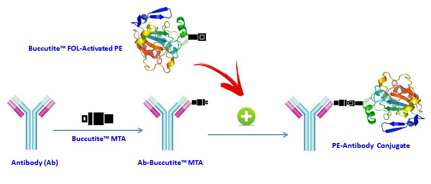
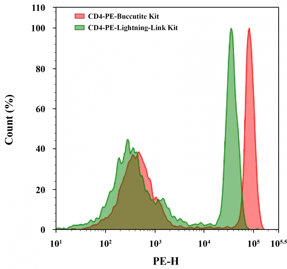
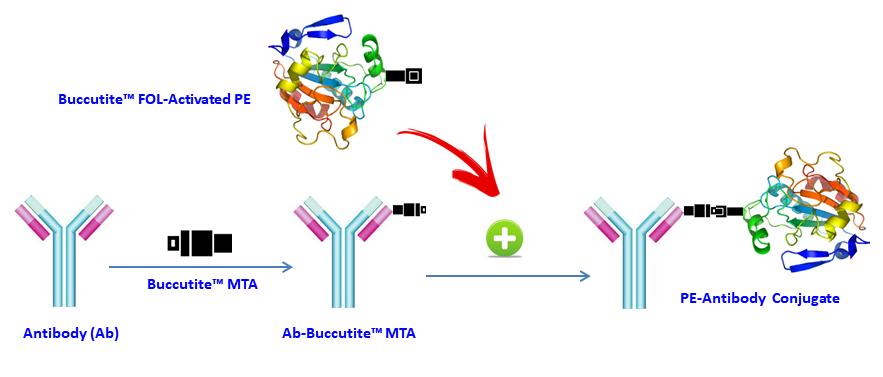
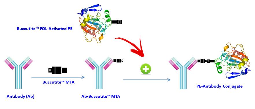
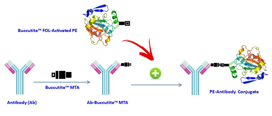
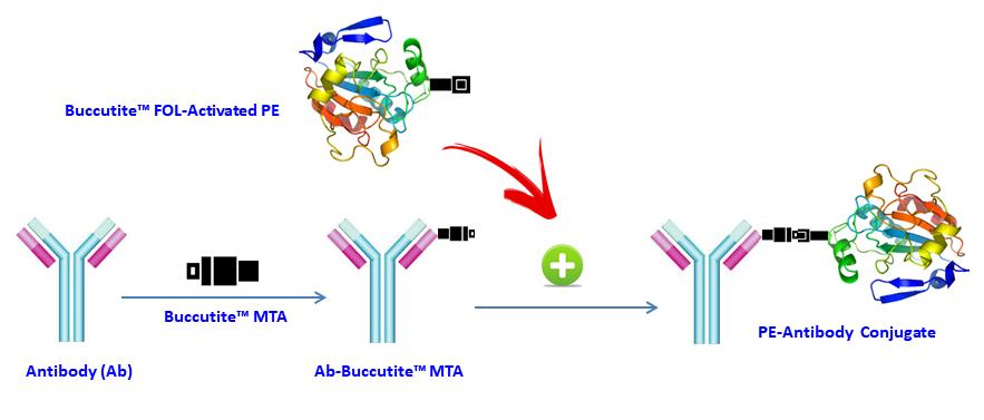
Buccutite RPE 抗体标记试剂盒提供了一种以微观方式标记抗体的便捷方法。RPE是具有565nm额定的激发波长和575nm的发射波长的橙色荧光蛋白。 ReadiLink RPE共轭试剂盒提供了一种简单方便的方法,灵活运用于你的抗体与视网膜色素上皮。共轭抗体可以在WB,ELISA和免疫组织化学应用中使用。本试剂盒足够用于2个标记反应,每次多50微克抗体。考虑PE(240 kDa)的大尺寸,抗体在标记反应中使用的量必须总是小于RPE的量。对于任何新的抗体试剂的佳比例,必须通过实验来确定,但50-60微克IgG抗体每100微克的RPE通常是佳结果。 60微克抗体约对应于一个抗体:1:1 RPE的摩尔比。Buccutite RPE抗体标记试剂盒是美国AAT Bioquest研发的产品。

点击查看光谱

实验方案

实验方案

操作步骤

1.运行Antibody-Buccutite MTA反应

1.1将抗体溶液直接加入到Buccutite MTA(组分B)的小瓶中,并通过反复移液几次将其充分混合或将小瓶涡旋几秒钟。

1.2将抗体-Buccutite MTA反应混合物在室温下保持30-60分钟。 注意:如果需要,抗体-Buccutite MTA反应混合物可以旋转或摇动更长时间。


2.制备抗体-PE缀合物

4.1通过向Buccutite FOL活化的PE(组分A)的小瓶中加入50μLddH2O制备Buccutite FOL活化的PE溶液,通过反复移液几次充分混合或将小瓶涡旋几秒钟。

4.2将整瓶Buccutite FOL活化的PE溶液混合到纯化的抗体-Buccutite MTA溶液(来自步骤纯化抗体-Buccutite MTA溶液)中,充分混合并在室温下旋转混合物1小时。

4.3抗体-PE结合物现在可以使用了。 注意:立即使用时,抗体-PE结合物需要用您选择的缓冲液稀释。 注意:对于长期储存,需要浓缩或冷冻干燥抗体-PE结合物溶液。


3.抗体-PE共轭物的储存

抗体缀合物应在载体抗体(例如0.1%牛血清白蛋白)存在下以> 0.5mg / mL储存。 当在2mM叠氮化钠存在下储存并避光时,抗体-PE缀合物溶液可以在4℃下储存两个月而没有显着变化。 对于更长时间的储存,可以将抗体-PE缀合物冻干并在≤-20℃下储存。


参考文献

Chromophore attachment to phycobiliprotein beta-subunits: phycocyanobilin:cysteine-beta84 phycobiliprotein lyase activity of CpeS-like protein from Anabaena Sp. PCC7120
Authors: Zhao KH, Su P, Li J, Tu JM, Zhou M, Bubenzer C, Scheer H.
Journal: J Biol Chem (2006): 8573

Excitation energy transfer from phycobiliprotein to chlorophyll d in intact cells of Acaryochloris marina studied by time- and wavelength-resolved fluorescence spectroscopy
Authors: Petrasek Z, Schmitt FJ, Theiss C, Huyer J, Chen M, Larkum A, Eichler HJ, Kemnitz K, Eckert HJ.
Journal: Photochem Photobiol Sci (2005): 1016

Single-molecule spectroscopy selectively probes donor and acceptor chromophores in the phycobiliprotein allophycocyanin
Authors: Loos D, Cotlet M, De Schryver F, Habuchi S, Hofkens J.
Journal: Biophys J (2004): 2598

Evaluation of Tolypothrix germplasm for phycobiliprotein content
Authors: Prasanna R, Prasanna BM, Mohammadi SA, Singh PK.
Journal: Folia Microbiol (Praha) (2003): 59

Isolation and characterisation of phycobiliprotein rich mutant of cyanobacterium Synechocystis sp
Authors: Prasanna R, Dhar DW, Dominic TK, Tiwari ON, Singh PK.
Journal: Acta Biol Hung (2003): 113

Co-ordinated expression of phycobiliprotein operons in the chromatically adapting cyanobacterium Calothrix PCC 7601: a role for RcaD and RcaG
Authors: Noubir S, Luque I, Ochoa de Alda JA, Perewoska I, Tandeau de Marsac N, Cobley JG, Houmard J.
Journal: Mol Microbiol (2002): 749

Phycobiliprotein genes of the marine photosynthetic prokaryote Prochlorococcus: evidence for rapid evolution of genetic heterogeneity
Authors: Ting CS, Rocap G, King J, Chisholm SW.
Journal: Microbiology (2001): 3171

Novel activity of a phycobiliprotein lyase: both the attachment of phycocyanobilin and the isomerization to phycoviolobilin are catalyzed by the proteins PecE and PecF encoded by the phycoerythrocyanin operon
Authors: Zhao KH, Deng MG, Zheng M, Zhou M, Parbel A, Storf M, Meyer M, Strohmann B, Scheer H.
Journal: FEBS Lett (2000): 9

Phycobiliprotein-Fab conjugates as probes for single particle fluorescence imaging
Authors: Triantafilou K, Triantafilou M, Wilson KM.
Journal: Cytometry (2000): 226

[Phycobiliprotein and fluorescence immunological assay]
Authors: Wu P.
Journal: Sheng Li Ke Xue Jin Zhan (2000): 82


相关产品

产品名称 货号
Buccutite RPE抗体标记试剂盒 标记25ug抗体 Cat#1312
Buccutite PerCP抗体标记试剂盒 标记25ug抗体 Cat#1353
Buccutite APC抗体标记试剂盒 标记100ug抗体 Cat#1311

Buccutite PE-Cy5.5抗体标记试剂盒 标记100ug抗体-AAT Bioquest荧光染料

上海金畔生物科技有限公司代理AAT Bioquest荧光染料全线产品,欢迎访问AAT Bioquest荧光染料官网了解更多信息。
Buccutite PE-Cy5.5抗体标记试剂盒 标记100ug抗体价格 7092
产品规格

2 Labelings

产品货号

Buccutite PE-Cy5.5抗体标记试剂盒 标记100ug抗体

产品参数
Ex (nm) 565 Em (nm) 671
分子量 溶剂 DMSO
存储条件 在2-8度冷藏保存, 避免光照
产品概述

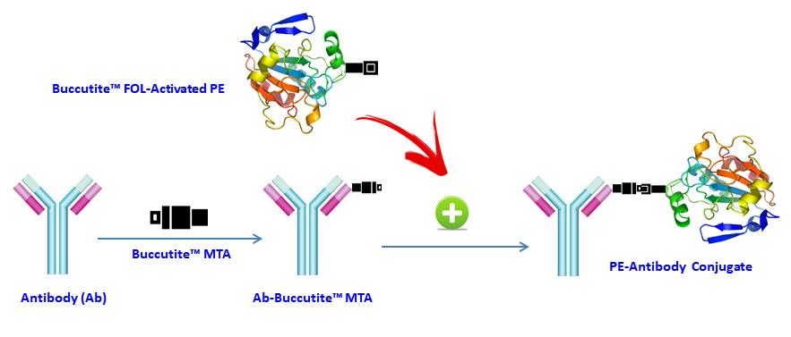
Buccutite PE-Cy5.5 抗体标记试剂盒提供了一种以微观方式标记抗体的便捷方法。PE-Cy5.5是流式细胞术中常用的颜色。其主要吸收峰位于565nm,发射峰位于~700nm。对于这种串联颜色,推荐使用682/33 nm和695/40 nm的滤光片组。 AAT Bioquest提供这种Buccutite 快速标记试剂盒,以促进PE-Cy5.5与抗体和其他蛋白质(如链霉抗生物素蛋白和其他二级试剂)的串联结合。 Buccutite PE-Cy5.5共轭试剂盒为您的抗体与PE结合提供了一种强大而方便的方法。该试剂盒包括活化的PE和反应缓冲液。缀合的抗体可用于WB,ELISA和IHC应用。该试剂盒足以进行2次标记反应,每次反应多100μg抗体。考虑到PE的大尺寸(240kDa),标记反应中使用的抗体量必须始终小于RPE的量。任何新抗体试剂的佳比例必须通过实验确定,但每100ug RPE 50-60ug IgG抗体通常可获得结果。我们的试剂盒提供预活化的PE-Cy5.5,以促进PE-Cy5.5串联结合抗体和其他蛋白质,如链霉抗生物素蛋白和其他二级试剂。我们的预活化PE-Cy5.5串联已准备好结合,产生比常规繁琐的基于SMCC的缀合化学高得多的产率。此外,我们的预活化PE-Cy5.5串联通过其蛋白质中丰富的氨基与蛋白质缀合,而SMCC化学靶向必须通过抗体还原而再生的硫醇基团。Buccutite PE-Cy5.5抗体标记试剂盒是美国AAT Bioquest研发的产品。

点击查看光谱

实验方案

实验方案

操作步骤

1.运行Antibody-Buccutite MTA反应

1.1将抗体溶液直接加入到Buccutite MTA(组分B)的小瓶中,并通过反复移液几次将其充分混合或将小瓶涡旋几秒钟。

1.2将抗体-Buccutite MTA反应混合物在室温下保持30-60分钟。 注意:如果需要,抗体-Buccutite MTA反应混合物可以旋转或摇动更长时间。


2.制备抗体-PE-Cy5.5缀合物

2.1通过向Buccutite FOL活化的PE-Cy5.5(组分A)的小瓶中加入50μLddH2O制备Buccutite FOL活化的PE溶液,通过反复移液几次充分混合或将小瓶涡旋几秒钟。

2.2将整瓶Buccutite FOL活化的PE-Cy5.5溶液混合到纯化的抗体-Buccutite MTA溶液(来自步骤纯化抗体-Buccutite MTA溶液)中,充分混合并在室温下旋转混合物1小时。

2.3抗体-PE-Cy5.5结合物现在可以使用了。 注意:立即使用时,抗体-PE-Cy5.5结合物需要用您选择的缓冲液稀释。 注意:对于长期储存,需要浓缩或冷冻干燥抗体-PE结合物溶液。


3.抗体-PE-Cy5.5共轭物的储存

抗体缀合物应在载体抗体(例如0.1%牛血清白蛋白)存在下以> 0.5mg / mL储存。 当在2mM叠氮化钠存在下储存并避光时,抗体-PE-Cy5.5缀合物溶液可以在4℃下储存两个月而没有显着变化。 对于更长时间的储存,可以将抗体-PE-Cy5.5缀合物冻干并在≤-20℃下储存。


参考文献

Chromophore attachment to phycobiliprotein beta-subunits: phycocyanobilin:cysteine-beta84 phycobiliprotein lyase activity of CpeS-like protein from Anabaena Sp. PCC7120
Authors: Zhao KH, Su P, Li J, Tu JM, Zhou M, Bubenzer C, Scheer H.
Journal: J Biol Chem (2006): 8573

Excitation energy transfer from phycobiliprotein to chlorophyll d in intact cells of Acaryochloris marina studied by time- and wavelength-resolved fluorescence spectroscopy
Authors: Petrasek Z, Schmitt FJ, Theiss C, Huyer J, Chen M, Larkum A, Eichler HJ, Kemnitz K, Eckert HJ.
Journal: Photochem Photobiol Sci (2005): 1016

Single-molecule spectroscopy selectively probes donor and acceptor chromophores in the phycobiliprotein allophycocyanin
Authors: Loos D, Cotlet M, De Schryver F, Habuchi S, Hofkens J.
Journal: Biophys J (2004): 2598

Evaluation of Tolypothrix germplasm for phycobiliprotein content
Authors: Prasanna R, Prasanna BM, Mohammadi SA, Singh PK.
Journal: Folia Microbiol (Praha) (2003): 59

Isolation and characterisation of phycobiliprotein rich mutant of cyanobacterium Synechocystis sp
Authors: Prasanna R, Dhar DW, Dominic TK, Tiwari ON, Singh PK.
Journal: Acta Biol Hung (2003): 113

Co-ordinated expression of phycobiliprotein operons in the chromatically adapting cyanobacterium Calothrix PCC 7601: a role for RcaD and RcaG
Authors: Noubir S, Luque I, Ochoa de Alda JA, Perewoska I, Tandeau de Marsac N, Cobley JG, Houmard J.
Journal: Mol Microbiol (2002): 749

Phycobiliprotein genes of the marine photosynthetic prokaryote Prochlorococcus: evidence for rapid evolution of genetic heterogeneity
Authors: Ting CS, Rocap G, King J, Chisholm SW.
Journal: Microbiology (2001): 3171

Novel activity of a phycobiliprotein lyase: both the attachment of phycocyanobilin and the isomerization to phycoviolobilin are catalyzed by the proteins PecE and PecF encoded by the phycoerythrocyanin operon
Authors: Zhao KH, Deng MG, Zheng M, Zhou M, Parbel A, Storf M, Meyer M, Strohmann B, Scheer H.
Journal: FEBS Lett (2000): 9

Phycobiliprotein-Fab conjugates as probes for single particle fluorescence imaging
Authors: Triantafilou K, Triantafilou M, Wilson KM.
Journal: Cytometry (2000): 226

[Phycobiliprotein and fluorescence immunological assay]
Authors: Wu P.
Journal: Sheng Li Ke Xue Jin Zhan (2000): 82


相关产品

产品名称 货号
Buccutite PE-Cy5.5抗体标记试剂盒 标记25ug抗体 Cat#1341
Buccutite APC-Cy5.5抗体标记试剂盒 标记100ug抗体 Cat#1320
Buccutite APC-Cy5.5抗体标记试剂盒 Cat#1350

Buccutite PE-Cy7抗体标记试剂盒 标记100ug抗体-AAT Bioquest荧光染料

上海金畔生物科技有限公司代理AAT Bioquest荧光染料全线产品,欢迎访问AAT Bioquest荧光染料官网了解更多信息。
Buccutite PE-Cy7抗体标记试剂盒 标记100ug抗体价格 7092
产品规格

2 Labelings

产品货号

Buccutite PE-Cy7抗体标记试剂盒 标记100ug抗体

产品参数
Ex (nm) 565 Em (nm) 778
分子量 溶剂
存储条件 在2-8度冷藏保存, 避免光照
产品概述

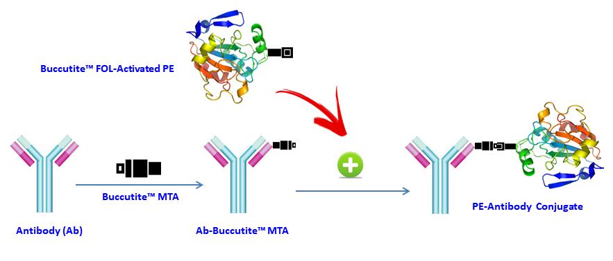
Buccutite PE-Cy7 抗体标记试剂盒提供了一种以微观方式标记抗体的便捷方法。PE-Cy7是流式细胞术中常用的颜色。其主要吸收峰位于565nm,发射峰位于~780nm。 AAT Bioquest提供这种Buccutite 快速标记试剂盒,以促进PE-Cy7串联结合抗体和其他蛋白质,如链霉抗生物素蛋白和其他二级试剂。 Buccutite PE-Cy7共轭试剂盒为您的抗体与PE结合提供了一种强大而方便的方法。该试剂盒包括活化的PE和反应缓冲液。缀合的抗体可用于WB,ELISA和IHC应用。该试剂盒足以进行2次标记反应,每次反应多100μg抗体。考虑到PE的大尺寸(240kDa),标记反应中使用的抗体量必须始终小于RPE的量。任何新抗体试剂的比例必须通过实验确定,但每100ug RPE 50-60ug IgG抗体通常可获得结果。我们的试剂盒提供预活化的PE-Cy7,以促进PE-Cy7串联结合抗体和其他蛋白质,如链霉抗生物素蛋白和其他二级试剂。我们的预活化PE-Cy7串联已准备好结合,产生比常规繁琐的基于SMCC的缀合化学高得多的产率。此外,我们的预活化PE-Cy7串联通过其蛋白质中丰富的氨基与蛋白质缀合,而SMCC化学靶向必须通过抗体还原而再生的硫醇基团。Buccutite PE-Cy7抗体标记试剂盒是美国AAT Bioquest研发的产品。

点击查看光谱

实验方案

实验方案

操作步骤

1.运行Antibody-Buccutite MTA反应

1.1将抗体溶液直接加入到Buccutite MTA(组分B)的小瓶中,并通过反复移液几次将其充分混合或将小瓶涡旋几秒钟。

1.2将抗体-Buccutite MTA反应混合物在室温下保持30-60分钟。 注意:如果需要,抗体-Buccutite MTA反应混合物可以旋转或摇动更长时间。


2.制备抗体-PE-Cy7缀合物

2.1通过向Buccutite FOL活化的PE-Cy7(组分A)的小瓶中加入50μLddH2O制备Buccutite FOL活化的PE溶液,通过反复移液几次充分混合或将小瓶涡旋几秒钟。

2.2将整瓶Buccutite FOL活化的PE-Cy7溶液混合到纯化的抗体-Buccutite MTA溶液(来自步骤纯化抗体-Buccutite MTA溶液)中,充分混合并在室温下旋转混合物1小时。

2.3抗体-PE-Cy7结合物现在可以使用了。 注意:立即使用时,抗体-PE-Cy7结合物需要用您选择的缓冲液稀释。 注意:对于长期储存,需要浓缩或冷冻干燥抗体-PE结合物溶液。


3.抗体-PE-Cy7共轭物的储存

抗体缀合物应在载体抗体(例如0.1%牛血清白蛋白)存在下以> 0.5mg / mL储存。 当在2mM叠氮化钠存在下储存并避光时,抗体-PE-Cy7缀合物溶液可以在4℃下储存两个月而没有显着变化。 对于更长时间的储存,可以将抗体-PE-Cy7缀合物冻干并在≤-20℃下储存。


参考文献

Chromophore attachment to phycobiliprotein beta-subunits: phycocyanobilin:cysteine-beta84 phycobiliprotein lyase activity of CpeS-like protein from Anabaena Sp. PCC7120
Authors: Zhao KH, Su P, Li J, Tu JM, Zhou M, Bubenzer C, Scheer H.
Journal: J Biol Chem (2006): 8573

Excitation energy transfer from phycobiliprotein to chlorophyll d in intact cells of Acaryochloris marina studied by time- and wavelength-resolved fluorescence spectroscopy
Authors: Petrasek Z, Schmitt FJ, Theiss C, Huyer J, Chen M, Larkum A, Eichler HJ, Kemnitz K, Eckert HJ.
Journal: Photochem Photobiol Sci (2005): 1016

Single-molecule spectroscopy selectively probes donor and acceptor chromophores in the phycobiliprotein allophycocyanin
Authors: Loos D, Cotlet M, De Schryver F, Habuchi S, Hofkens J.
Journal: Biophys J (2004): 2598

Evaluation of Tolypothrix germplasm for phycobiliprotein content
Authors: Prasanna R, Prasanna BM, Mohammadi SA, Singh PK.
Journal: Folia Microbiol (Praha) (2003): 59

Isolation and characterisation of phycobiliprotein rich mutant of cyanobacterium Synechocystis sp
Authors: Prasanna R, Dhar DW, Dominic TK, Tiwari ON, Singh PK.
Journal: Acta Biol Hung (2003): 113

Co-ordinated expression of phycobiliprotein operons in the chromatically adapting cyanobacterium Calothrix PCC 7601: a role for RcaD and RcaG
Authors: Noubir S, Luque I, Ochoa de Alda JA, Perewoska I, Tandeau de Marsac N, Cobley JG, Houmard J.
Journal: Mol Microbiol (2002): 749

Phycobiliprotein genes of the marine photosynthetic prokaryote Prochlorococcus: evidence for rapid evolution of genetic heterogeneity
Authors: Ting CS, Rocap G, King J, Chisholm SW.
Journal: Microbiology (2001): 3171

Novel activity of a phycobiliprotein lyase: both the attachment of phycocyanobilin and the isomerization to phycoviolobilin are catalyzed by the proteins PecE and PecF encoded by the phycoerythrocyanin operon
Authors: Zhao KH, Deng MG, Zheng M, Zhou M, Parbel A, Storf M, Meyer M, Strohmann B, Scheer H.
Journal: FEBS Lett (2000): 9

Phycobiliprotein-Fab conjugates as probes for single particle fluorescence imaging
Authors: Triantafilou K, Triantafilou M, Wilson KM.
Journal: Cytometry (2000): 226

[Phycobiliprotein and fluorescence immunological assay]
Authors: Wu P.
Journal: Sheng Li Ke Xue Jin Zhan (2000): 82


相关产品

产品名称 货号
Buccutite PE-Cy5.5抗体标记试剂盒 标记25ug抗体 Cat#1341
Buccutite APC-Cy5.5抗体标记试剂盒 标记100ug抗体 Cat#1320
Buccutite APC-Cy5.5抗体标记试剂盒 Cat#1350

Buccutite PE-Texas Red抗体标记试剂盒 标记100ug抗体-AAT Bioquest荧光染料

上海金畔生物科技有限公司代理AAT Bioquest荧光染料全线产品,欢迎访问AAT Bioquest荧光染料官网了解更多信息。
Buccutite PE-Texas Red抗体标记试剂盒 标记100ug抗体价格 7092
产品规格

2 Labelings

产品货号

Buccutite PE-Texas Red抗体标记试剂盒 标记100ug抗体

产品参数
Ex (nm) 565 Em (nm) 613
分子量 溶剂 DMSO
存储条件 在2-8度冷藏保存, 避免光照
产品概述

Buccutite PE-Texas Red 抗体标记试剂盒提供了一种以微观方式标记抗体的便捷方法。PE-Texas Red是流式细胞仪中常用的颜色。其主要吸收峰位于565nm,发射峰位于600nm。 AAT Bioquest提供这种Buccutite 快速标记试剂盒,以促进PE-Texas Red串联结合抗体和其他蛋白质,如链霉抗生物素蛋白和其他二级试剂。 Buccutite PE-Texas Red Conjugation Kit为您的抗体与PE结合提供了一种强大而方便的方法。该试剂盒包括活化的PE和反应缓冲液。缀合的抗体可用于WB,ELISA和IHC应用。该试剂盒足以进行2次标记反应,每次反应多100μg抗体。考虑到PE的大尺寸(240kDa),标记反应中使用的抗体量必须始终小于RPE的量。任何新抗体试剂的比例必须通过实验确定,但每100ug RPE 50-60ug IgG抗体通常可获得结果。我们的试剂盒提供预活化的PE-Texas Red,以促进PE-Texas Red串联结合抗体和其他蛋白质,如链霉抗生物素蛋白和其他二级试剂。我们的预活化PE-Texas Red tandem准备结合,产生比常规繁琐的基于SMCC的缀合化学高得多的产率。此外,我们的预活化PE-Texas Red串联通过其蛋白质丰富的氨基与蛋白质缀合,而SMCC化学靶向必须通过抗体还原而再生的硫醇基团。Buccutite PE-Texas Red抗体标记试剂盒是美国AAT Bioquest研发的产品。

点击查看光谱

实验方案

实验方案

操作步骤

1.运行Antibody-Buccutite MTA反应

1.1将抗体溶液直接加入到Buccutite MTA(组分B)的小瓶中,并通过反复移液几次将其充分混合或将小瓶涡旋几秒钟。

1.2将抗体-Buccutite MTA反应混合物在室温下保持30-60分钟。 注意:如果需要,抗体-Buccutite MTA反应混合物可以旋转或摇动更长时间。


2.制备抗体-PE-Texas Red缀合物

2.1通过向Buccutite FOL活化的PE-Texas Red(组分A)的小瓶中加入50μLddH2O制备Buccutite FOL活化的PE溶液,通过反复移液几次充分混合或将小瓶涡旋几秒钟。

2.2将整瓶Buccutite FOL活化的PE-Texas Red溶液混合到纯化的抗体-Buccutite MTA溶液(来自步骤纯化抗体-Buccutite MTA溶液)中,充分混合并在室温下旋转混合物1小时。

2.3抗体-PE-Texas Red结合物现在可以使用了。 注意:立即使用时,抗体-PE-Texas Red结合物需要用您选择的缓冲液稀释。 注意:对于长期储存,需要浓缩或冷冻干燥抗体-PE结合物溶液。


3.抗体-PE-Texas Red共轭物的储存

抗体缀合物应在载体抗体(例如0.1%牛血清白蛋白)存在下以> 0.5mg / mL储存。 当在2mM叠氮化钠存在下储存并避光时,抗体-PE-Texas Red缀合物溶液可以在4℃下储存两个月而没有显着变化。 对于更长时间的储存,可以将抗体-PE-Texas Red缀合物冻干并在≤-20℃下储存。


参考文献

Chromophore attachment to phycobiliprotein beta-subunits: phycocyanobilin:cysteine-beta84 phycobiliprotein lyase activity of CpeS-like protein from Anabaena Sp. PCC7120
Authors: Zhao KH, Su P, Li J, Tu JM, Zhou M, Bubenzer C, Scheer H.
Journal: J Biol Chem (2006): 8573

Excitation energy transfer from phycobiliprotein to chlorophyll d in intact cells of Acaryochloris marina studied by time- and wavelength-resolved fluorescence spectroscopy
Authors: Petrasek Z, Schmitt FJ, Theiss C, Huyer J, Chen M, Larkum A, Eichler HJ, Kemnitz K, Eckert HJ.
Journal: Photochem Photobiol Sci (2005): 1016

Single-molecule spectroscopy selectively probes donor and acceptor chromophores in the phycobiliprotein allophycocyanin
Authors: Loos D, Cotlet M, De Schryver F, Habuchi S, Hofkens J.
Journal: Biophys J (2004): 2598

Evaluation of Tolypothrix germplasm for phycobiliprotein content
Authors: Prasanna R, Prasanna BM, Mohammadi SA, Singh PK.
Journal: Folia Microbiol (Praha) (2003): 59

Isolation and characterisation of phycobiliprotein rich mutant of cyanobacterium Synechocystis sp
Authors: Prasanna R, Dhar DW, Dominic TK, Tiwari ON, Singh PK.
Journal: Acta Biol Hung (2003): 113

Co-ordinated expression of phycobiliprotein operons in the chromatically adapting cyanobacterium Calothrix PCC 7601: a role for RcaD and RcaG
Authors: Noubir S, Luque I, Ochoa de Alda JA, Perewoska I, Tandeau de Marsac N, Cobley JG, Houmard J.
Journal: Mol Microbiol (2002): 749

Phycobiliprotein genes of the marine photosynthetic prokaryote Prochlorococcus: evidence for rapid evolution of genetic heterogeneity
Authors: Ting CS, Rocap G, King J, Chisholm SW.
Journal: Microbiology (2001): 3171

Novel activity of a phycobiliprotein lyase: both the attachment of phycocyanobilin and the isomerization to phycoviolobilin are catalyzed by the proteins PecE and PecF encoded by the phycoerythrocyanin operon
Authors: Zhao KH, Deng MG, Zheng M, Zhou M, Parbel A, Storf M, Meyer M, Strohmann B, Scheer H.
Journal: FEBS Lett (2000): 9

Phycobiliprotein-Fab conjugates as probes for single particle fluorescence imaging
Authors: Triantafilou K, Triantafilou M, Wilson KM.
Journal: Cytometry (2000): 226

[Phycobiliprotein and fluorescence immunological assay]
Authors: Wu P.
Journal: Sheng Li Ke Xue Jin Zhan (2000): 82


相关产品

产品名称 货号
Buccutite PE-Cy5.5抗体标记试剂盒 标记25ug抗体 Cat#1341
Buccutite APC-Cy5.5抗体标记试剂盒 标记100ug抗体 Cat#1320
Buccutite APC-Cy5.5抗体标记试剂盒 Cat#1350

Buccutite PE-Cy5抗体标记试剂盒 标记100ug抗体-AAT Bioquest荧光染料

上海金畔生物科技有限公司代理AAT Bioquest荧光染料全线产品,欢迎访问AAT Bioquest荧光染料官网了解更多信息。
Buccutite PE-Cy5抗体标记试剂盒 标记100ug抗体价格 7092
产品规格

2 Labelings

产品货号

Buccutite PE-Cy5抗体标记试剂盒 标记100ug抗体

产品参数
Ex (nm) 565 Em (nm) 666
分子量 溶剂 DMSO
存储条件 在2-8度冷藏保存, 避免光照
产品概述

Buccutite PE-Cy5抗体标记试剂盒提供了一种以微观方式标记抗体的便捷方法。PE-Cy5是流式细胞术中流行的一种颜色。其主要吸收峰位于565 nm,发射峰位于674 nm。对于这种串联颜色,推荐使用682/33 nm和695/40 nm的滤光片组。 AAT Bioquest提供这种Buccutite 快速标记试剂盒,以促进PE-Cy5串联结合抗体和其他蛋白质,如链霉抗生物素蛋白和其他二级试剂。 Buccutite PE-Cy5缀合试剂盒提供了一种稳定且便捷的方法来将您的抗体与PE结合。该试剂盒包括活化的PE和反应缓冲液。缀合的抗体可用于WB,ELISA和IHC应用。该试剂盒足以进行2次标记反应,每次反应多100μg抗体。考虑到PE的大尺寸(240kDa),标记反应中使用的抗体量必须小于RPE的量。任何新抗体试剂的比例必须通过实验确定,但每100ug RPE 50-60ug IgG抗体通常可获得结果。我们的试剂盒提供预活化的PE-Cy5,以促进PE-Cy5串联结合抗体和其他蛋白质,如链霉抗生物素蛋白和其他二级试剂。我们预活化的PE-Cy5串联准备结合,比常规繁琐的基于SMCC的结合化学提供更高的产量。此外,我们的预活化PE-Cy5串联通过其蛋白质丰富的氨基与蛋白质缀合,而SMCC化学靶向必须通过抗体还原而再生的硫醇基团。 Buccutite PE-Cy5抗体标记试剂盒是美国AAT Bioquest研发的产品。

点击查看光谱

实验方案

实验方案

操作步骤

1.运行Antibody-Buccutite MTA反应

1.1将抗体溶液直接加入到Buccutite MTA(组分B)的小瓶中,并通过反复移液几次将其充分混合或将小瓶涡旋几秒钟。

1.2将抗体-Buccutite MTA反应混合物在室温下保持30-60分钟。 注意:如果需要,抗体-Buccutite MTA反应混合物可以旋转或摇动更长时间。


2.制备抗体-PE-Cy5缀合物

2.1通过向Buccutite FOL活化的PE-Cy5(组分A)的小瓶中加入50μLddH2O制备Buccutite FOL活化的PE-Cy5溶液,通过反复移液几次充分混合或将小瓶涡旋几秒钟。

2.2将整瓶Buccutite FOL活化的PE-Cy5溶液混合到纯化的抗体-Buccutite MTA溶液(来自步骤纯化抗体-Buccutite MTA溶液)中,充分混合并在室温下旋转混合物1小时。

2.3抗体-PE-Cy5结合物现在可以使用了。 注意:立即使用时,抗体-PE-Cy5结合物需要用您选择的缓冲液稀释。 注意:对于长期储存,需要浓缩或冷冻干燥抗体-PE-Cy5结合物溶液。


3.抗体-PE-Cy5共轭物的储存

抗体缀合物应在载体抗体(例如0.1%牛血清白蛋白)存在下以> 0.5mg / mL储存。 当在2mM叠氮化钠存在下储存并避光时,抗体-PE-Cy5缀合物溶液可以在4℃下储存两个月而没有显着变化。 对于更长时间的储存,可以将抗体-PE-Cy5缀合物冻干并在≤-20℃下储存。


参考文献

Chromophore attachment to phycobiliprotein beta-subunits: phycocyanobilin:cysteine-beta84 phycobiliprotein lyase activity of CpeS-like protein from Anabaena Sp. PCC7120
Authors: Zhao KH, Su P, Li J, Tu JM, Zhou M, Bubenzer C, Scheer H.
Journal: J Biol Chem (2006): 8573

Excitation energy transfer from phycobiliprotein to chlorophyll d in intact cells of Acaryochloris marina studied by time- and wavelength-resolved fluorescence spectroscopy
Authors: Petrasek Z, Schmitt FJ, Theiss C, Huyer J, Chen M, Larkum A, Eichler HJ, Kemnitz K, Eckert HJ.
Journal: Photochem Photobiol Sci (2005): 1016

Single-molecule spectroscopy selectively probes donor and acceptor chromophores in the phycobiliprotein allophycocyanin
Authors: Loos D, Cotlet M, De Schryver F, Habuchi S, Hofkens J.
Journal: Biophys J (2004): 2598

Evaluation of Tolypothrix germplasm for phycobiliprotein content
Authors: Prasanna R, Prasanna BM, Mohammadi SA, Singh PK.
Journal: Folia Microbiol (Praha) (2003): 59

Isolation and characterisation of phycobiliprotein rich mutant of cyanobacterium Synechocystis sp
Authors: Prasanna R, Dhar DW, Dominic TK, Tiwari ON, Singh PK.
Journal: Acta Biol Hung (2003): 113

Co-ordinated expression of phycobiliprotein operons in the chromatically adapting cyanobacterium Calothrix PCC 7601: a role for RcaD and RcaG
Authors: Noubir S, Luque I, Ochoa de Alda JA, Perewoska I, Tandeau de Marsac N, Cobley JG, Houmard J.
Journal: Mol Microbiol (2002): 749

Phycobiliprotein genes of the marine photosynthetic prokaryote Prochlorococcus: evidence for rapid evolution of genetic heterogeneity
Authors: Ting CS, Rocap G, King J, Chisholm SW.
Journal: Microbiology (2001): 3171

Novel activity of a phycobiliprotein lyase: both the attachment of phycocyanobilin and the isomerization to phycoviolobilin are catalyzed by the proteins PecE and PecF encoded by the phycoerythrocyanin operon
Authors: Zhao KH, Deng MG, Zheng M, Zhou M, Parbel A, Storf M, Meyer M, Strohmann B, Scheer H.
Journal: FEBS Lett (2000): 9

Phycobiliprotein-Fab conjugates as probes for single particle fluorescence imaging
Authors: Triantafilou K, Triantafilou M, Wilson KM.
Journal: Cytometry (2000): 226

[Phycobiliprotein and fluorescence immunological assay]
Authors: Wu P.
Journal: Sheng Li Ke Xue Jin Zhan (2000): 82


相关产品

产品名称 货号
Buccutite PE-Cy5抗体标记试剂盒 标记25ug抗体 Cat#1340
Buccutite APC-Cy7抗体标记试剂盒 标记100ug抗体 Cat#1321
Buccutite APC-Cy7抗体标记试剂盒 标记25ug抗体 Cat#1351

Buccutite PE-Cy5抗体标记试剂盒 标记25ug抗体-AAT Bioquest荧光染料

上海金畔生物科技有限公司代理AAT Bioquest荧光染料全线产品,欢迎访问AAT Bioquest荧光染料官网了解更多信息。
Buccutite PE-Cy5抗体标记试剂盒 标记25ug抗体价格 4245
产品规格

2 Labelings

产品货号

Buccutite PE-Cy5抗体标记试剂盒 标记25ug抗体

产品参数
Ex (nm) 565 Em (nm) 666
分子量 溶剂 DMSO
存储条件 在2-8度冷藏保存, 避免光照
产品概述

Buccutite PE-Cy5抗体标记试剂盒提供了一种以微观方式标记抗体的便捷方法。PE-Cy5是流式细胞术中常用的颜色。其主要吸收峰位于565nm,发射峰位于674nm。对于这种串联颜色,建议使用682/33 nm和695/40 nm的滤光片组。 AAT Bioquest提供这种Buccutite 快速标记试剂盒,以促进PE-Cy5串联结合抗体和其他蛋白质,如链霉抗生物素蛋白和其他二级试剂。 Buccutite PE-Cy5共轭试剂盒为您的抗体与PE结合提供了一种强大而方便的方法。该试剂盒包括预活化的PE和反应缓冲液。整个过程只需要两次简单的混合,无需进一步纯化。缀合的抗体可用于WB,ELISA和IHC应用。该试剂盒足以进行2次标记反应,每次反应 多25μg抗体。考虑到PE的大尺寸(240kDa),标记反应中使用的抗体量必须小于PE的量。任何新抗体试剂的 佳比例必须通过实验确定,但每50μgPE的25μgIgG抗体通常可获得 佳结果。我们的试剂盒提供预活化的PE-Cy5,以促进PE-Cy5串联结合抗体和其他蛋白质,如链霉抗生物素蛋白和其他二级试剂。我们的预活化PE-Cy5串联已准备好结合,产生比常规繁琐的基于SMCC的缀合化学高得多的产率。此外,我们的预活化PE-Cy5串联通过其蛋白质中丰富的氨基与蛋白质缀合,而SMCC化学靶向必须通过抗体还原而再生的硫醇基团。Buccutite PE-Cy5抗体标记试剂盒是美国AAT Bioquest研发的产品。

点击查看光谱

实验方案

实验方案

操作步骤

1.运行Antibody-Buccutite MTA反应

1.1将抗体溶液直接加入到Buccutite MTA(组分B)的小瓶中,并通过反复移液几次将其充分混合或将小瓶涡旋几秒钟。

1.2将抗体-Buccutite MTA反应混合物在室温下保持30-60分钟。 注意:如果需要,抗体-Buccutite MTA反应混合物可以旋转或摇动更长时间。


2.制备抗体-PE-Cy5缀合物

2.1通过向Buccutite FOL活化的PE-Cy5(组分A)的小瓶中加入50μLddH2O制备Buccutite FOL活化的PE-Cy5溶液,通过反复移液几次充分混合或将小瓶涡旋几秒钟。

2.2将整瓶Buccutite FOL活化的PE-Cy5溶液混合到纯化的抗体-Buccutite MTA溶液(来自步骤纯化抗体-Buccutite MTA溶液)中,充分混合并在室温下旋转混合物1小时。

2.3抗体-PE-Cy5结合物现在可以使用了。 注意:立即使用时,抗体-PE-Cy5结合物需要用您选择的缓冲液稀释。 注意:对于长期储存,需要浓缩或冷冻干燥抗体-PE-Cy5结合物溶液。


3.抗体-PE-Cy5共轭物的储存

抗体缀合物应在载体抗体(例如0.1%牛血清白蛋白)存在下以> 0.5mg / mL储存。 当在2mM叠氮化钠存在下储存并避光时,抗体-PE-Cy5缀合物溶液可以在4℃下储存两个月而没有显着变化。 对于更长时间的储存,可以将抗体-PE-Cy5缀合物冻干并在≤-20℃下储存。


参考文献

Chromophore attachment to phycobiliprotein beta-subunits: phycocyanobilin:cysteine-beta84 phycobiliprotein lyase activity of CpeS-like protein from Anabaena Sp. PCC7120
Authors: Zhao KH, Su P, Li J, Tu JM, Zhou M, Bubenzer C, Scheer H.
Journal: J Biol Chem (2006): 8573

Excitation energy transfer from phycobiliprotein to chlorophyll d in intact cells of Acaryochloris marina studied by time- and wavelength-resolved fluorescence spectroscopy
Authors: Petrasek Z, Schmitt FJ, Theiss C, Huyer J, Chen M, Larkum A, Eichler HJ, Kemnitz K, Eckert HJ.
Journal: Photochem Photobiol Sci (2005): 1016

Single-molecule spectroscopy selectively probes donor and acceptor chromophores in the phycobiliprotein allophycocyanin
Authors: Loos D, Cotlet M, De Schryver F, Habuchi S, Hofkens J.
Journal: Biophys J (2004): 2598

Evaluation of Tolypothrix germplasm for phycobiliprotein content
Authors: Prasanna R, Prasanna BM, Mohammadi SA, Singh PK.
Journal: Folia Microbiol (Praha) (2003): 59

Isolation and characterisation of phycobiliprotein rich mutant of cyanobacterium Synechocystis sp
Authors: Prasanna R, Dhar DW, Dominic TK, Tiwari ON, Singh PK.
Journal: Acta Biol Hung (2003): 113

Co-ordinated expression of phycobiliprotein operons in the chromatically adapting cyanobacterium Calothrix PCC 7601: a role for RcaD and RcaG
Authors: Noubir S, Luque I, Ochoa de Alda JA, Perewoska I, Tandeau de Marsac N, Cobley JG, Houmard J.
Journal: Mol Microbiol (2002): 749

Phycobiliprotein genes of the marine photosynthetic prokaryote Prochlorococcus: evidence for rapid evolution of genetic heterogeneity
Authors: Ting CS, Rocap G, King J, Chisholm SW.
Journal: Microbiology (2001): 3171

Novel activity of a phycobiliprotein lyase: both the attachment of phycocyanobilin and the isomerization to phycoviolobilin are catalyzed by the proteins PecE and PecF encoded by the phycoerythrocyanin operon
Authors: Zhao KH, Deng MG, Zheng M, Zhou M, Parbel A, Storf M, Meyer M, Strohmann B, Scheer H.
Journal: FEBS Lett (2000): 9

Phycobiliprotein-Fab conjugates as probes for single particle fluorescence imaging
Authors: Triantafilou K, Triantafilou M, Wilson KM.
Journal: Cytometry (2000): 226

[Phycobiliprotein and fluorescence immunological assay]
Authors: Wu P.
Journal: Sheng Li Ke Xue Jin Zhan (2000): 82


相关产品

产品名称 货号
Buccutite PE-Cy5抗体标记试剂盒 标记100ug抗体 Cat#1322
Buccutite APC-Cy7抗体标记试剂盒 标记100ug抗体 Cat#1321
Buccutite APC-Cy7抗体标记试剂盒 标记25ug抗体 Cat#1351

Buccutite PE-Cy5.5抗体标记试剂盒 标记25ug抗体-AAT Bioquest荧光染料

上海金畔生物科技有限公司代理AAT Bioquest荧光染料全线产品,欢迎访问AAT Bioquest荧光染料官网了解更多信息。
Buccutite PE-Cy5.5抗体标记试剂盒 标记25ug抗体价格 4245
产品规格

2 Labelings

产品货号

Buccutite PE-Cy5.5抗体标记试剂盒 标记25ug抗体

产品参数
Ex (nm) 565 Em (nm) 671
分子量 溶剂 DMSO
存储条件 在2-8度冷藏保存, 避免光照
产品概述

Buccutite PE-Cy5.5抗体标记试剂盒提供了一种以微观方式标记抗体的便捷方法。PE-Cy5.5是流式细胞仪中常用的颜色。其主要吸收峰位于565nm,发射峰位于~700nm。对于这种串联颜色,推荐使用682/33 nm和695/40 nm的滤光片组。 AAT Bioquest提供这种Buccutite 快速标记试剂盒,以促进PE-Cy5.5与抗体和其他蛋白质(如链霉抗生物素蛋白和其他二级试剂)的串联结合。 Buccutite PE-Cy5.5共轭试剂盒为您的抗体与PE结合提供了一种强大而方便的方法。该试剂盒包括预活化的PE和反应缓冲液。整个过程仅需要两次简单的混合,无需进一步纯化。缀合的抗体可用于WB,ELISA和IHC应用。该试剂盒足以进行2次标记反应,每次反应多25μg抗体。考虑到PE的大尺寸(240kDa),标记反应中使用的抗体量必须小于PE的量。任何新抗体试剂的比例必须通过实验确定,但每50μgPE使用25μgIgG抗体通常可获得结果。我们的试剂盒提供预活化的PE-Cy5.5,以促进PE-Cy5.5串联结合抗体和其他蛋白质,如链霉抗生物素蛋白和其他二级试剂。我们的预活化PE-Cy5.5串联已准备好结合,产生比常规繁琐的基于SMCC的缀合化学高得多的产率。此外,我们的预活化PE-Cy5.5串联通过其蛋白质丰富的氨基与蛋白质缀合,而SMCC化学靶向必须通过抗体还原而再生的硫醇基团。Buccutite PE-Cy5.5抗体标记试剂盒是美国AAT Bioquest研发的产品。

点击查看光谱

实验方案

实验方案

操作步骤

1.运行Antibody-Buccutite MTA反应

1.1将抗体溶液直接加入到Buccutite MTA(组分B)的小瓶中,并通过反复移液几次将其充分混合或将小瓶涡旋几秒钟。

1.2将抗体-Buccutite MTA反应混合物在室温下保持30-60分钟。 注意:如果需要,抗体-Buccutite MTA反应混合物可以旋转或摇动更长时间。


2.制备抗体-PE-Cy5.5缀合物

2.1通过向Buccutite FOL活化的PE-Cy5.5(组分A)的小瓶中加入50μLddH2O制备Buccutite FOL活化的PE溶液,通过反复移液几次充分混合或将小瓶涡旋几秒钟。

2.2将整瓶Buccutite FOL活化的PE-Cy5.5溶液混合到纯化的抗体-Buccutite MTA溶液(来自步骤纯化抗体-Buccutite MTA溶液)中,充分混合并在室温下旋转混合物1小时。

2.3抗体-PE-Cy5.5结合物现在可以使用了。 注意:立即使用时,抗体-PE-Cy5.5结合物需要用您选择的缓冲液稀释。 注意:对于长期储存,需要浓缩或冷冻干燥抗体-PE结合物溶液。


3.抗体-PE-Cy5.5共轭物的储存

抗体缀合物应在载体抗体(例如0.1%牛血清白蛋白)存在下以> 0.5mg / mL储存。 当在2mM叠氮化钠存在下储存并避光时,抗体-PE-Cy5.5缀合物溶液可以在4℃下储存两个月而没有显着变化。 对于更长时间的储存,可以将抗体-PE-Cy5.5缀合物冻干并在≤-20℃下储存。


参考文献

Chromophore attachment to phycobiliprotein beta-subunits: phycocyanobilin:cysteine-beta84 phycobiliprotein lyase activity of CpeS-like protein from Anabaena Sp. PCC7120
Authors: Zhao KH, Su P, Li J, Tu JM, Zhou M, Bubenzer C, Scheer H.
Journal: J Biol Chem (2006): 8573

Excitation energy transfer from phycobiliprotein to chlorophyll d in intact cells of Acaryochloris marina studied by time- and wavelength-resolved fluorescence spectroscopy
Authors: Petrasek Z, Schmitt FJ, Theiss C, Huyer J, Chen M, Larkum A, Eichler HJ, Kemnitz K, Eckert HJ.
Journal: Photochem Photobiol Sci (2005): 1016

Single-molecule spectroscopy selectively probes donor and acceptor chromophores in the phycobiliprotein allophycocyanin
Authors: Loos D, Cotlet M, De Schryver F, Habuchi S, Hofkens J.
Journal: Biophys J (2004): 2598

Evaluation of Tolypothrix germplasm for phycobiliprotein content
Authors: Prasanna R, Prasanna BM, Mohammadi SA, Singh PK.
Journal: Folia Microbiol (Praha) (2003): 59

Isolation and characterisation of phycobiliprotein rich mutant of cyanobacterium Synechocystis sp
Authors: Prasanna R, Dhar DW, Dominic TK, Tiwari ON, Singh PK.
Journal: Acta Biol Hung (2003): 113

Co-ordinated expression of phycobiliprotein operons in the chromatically adapting cyanobacterium Calothrix PCC 7601: a role for RcaD and RcaG
Authors: Noubir S, Luque I, Ochoa de Alda JA, Perewoska I, Tandeau de Marsac N, Cobley JG, Houmard J.
Journal: Mol Microbiol (2002): 749

Phycobiliprotein genes of the marine photosynthetic prokaryote Prochlorococcus: evidence for rapid evolution of genetic heterogeneity
Authors: Ting CS, Rocap G, King J, Chisholm SW.
Journal: Microbiology (2001): 3171

Novel activity of a phycobiliprotein lyase: both the attachment of phycocyanobilin and the isomerization to phycoviolobilin are catalyzed by the proteins PecE and PecF encoded by the phycoerythrocyanin operon
Authors: Zhao KH, Deng MG, Zheng M, Zhou M, Parbel A, Storf M, Meyer M, Strohmann B, Scheer H.
Journal: FEBS Lett (2000): 9

Phycobiliprotein-Fab conjugates as probes for single particle fluorescence imaging
Authors: Triantafilou K, Triantafilou M, Wilson KM.
Journal: Cytometry (2000): 226

[Phycobiliprotein and fluorescence immunological assay]
Authors: Wu P.
Journal: Sheng Li Ke Xue Jin Zhan (2000): 82


相关产品

产品名称 货号
Buccutite PE-Cy5.5抗体标记试剂盒 标记100ug抗体 Cat#1316
Buccutite APC-Cy5.5抗体标记试剂盒 标记100ug抗体 Cat#1320
Buccutite APC-Cy5.5抗体标记试剂盒 Cat#1350