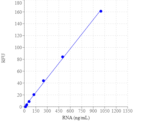
StrandBrite 绿色RNA定量试剂*200X DMSO溶解-AAT Bioquest荧光染料

上海金畔生物科技有限公司代理AAT Bioquest荧光染料全线产品,欢迎访问AAT Bioquest荧光染料官网了解更多信息。
StrandBrite 绿色RNA定量试剂*200X DMSO溶解 价格 4245
产品规格

1 ml

产品货号

StrandBrite 绿色RNA定量试剂*200X DMSO溶解

产品参数
Ex (nm) 509 Em (nm) 527
分子量 N/A 溶剂 DMSO
存储条件 在零下15度以下保存, 避免光照
产品概述

StrandBrite 绿色RNA定量试剂是美国AAT Bioquest生产的用于定量RNA的试剂,检测和定量少量RNA对于多种分子生物学程序极为重要,例如在进行Northern blot分析,S1核酸酶测定,RNase保护测定,cDNA文库制备,反向操作之前,测量体外转录RNA的产量和测量RNA浓度。转录PCR和差异展示PCR。测量核酸浓度常用的技术是测定260 nm的吸光度。基于吸收率的方法的主要缺点是蛋白质,游离核苷酸和其他紫外线吸收化合物引起的干扰。使用敏感的荧光核酸染色剂可缓解此干扰问题。StrandBrite RNA定量试剂是一种超灵敏的荧光核酸染料,用于定量溶液中的RNA。StrandBrite RNA定量试剂可通过荧光酶标仪或荧光计检测低至5 ng / mL的RNA。金畔生物是AAT Bioquest的中国代理商,为您提供优质的StrandBrite 绿色RNA定量试剂。 


适用仪器


荧光酶标仪  
Ex: 490 nm
Em: 525 nm
Cutoff: 515 nm
推荐孔板: 纯黑色孔板
   
实验方案

样品实验方案

溶液配制

工作溶液配制

1.准备StrandBrite 工作溶液:

1.1  准备的水溶液工作溶液StrandBrite 绿色通过在TE浓缩DMSO溶液的200倍稀释液(在DEPC的10mM的Tris-HCl,1mM的EDTA,pH 7.5中经处理的水)。例如,添加50μL StrandBrite 绿色 至10mL TE缓冲液,以制备足够的工作溶液在200至100个测定样品 μ L终体积。通过用箔纸覆盖或将其置于黑暗中来保护工作溶液免受光照。

注意1:我们建议在塑料容器中而不是玻璃中制备此溶液,因为染料可能会吸附到玻璃表面。

注意2:为获得佳结果,应在准备后的几个小时内使用此溶液。


标准液配制:

1.准备RNA标准液的系列稀释液(0到1µg / mL ):

1.1准备一个100 μ 克/ mL储备在RNA的溶液DEPC处理过的水。

1.2添加10μL 100 μ 克/毫升RNA储备溶液(来自步骤2.1) ,以490 μ 大号测定缓冲液(成分B)为具有2 微克/ mL的RNA溶液,然后执行1:2系列稀释来获得1000, 500、250、125、62.5、31.3、15.6和0 ng / mL。

1.3如表1和表2所述,将RNA标准品和含RNA 的测试样品添加到96孔黑色固体微孔板中。


表1黑色固体96孔微孔板中RNA标准品和测试样品的布局

BL BL TS TS
RS1 RS1
RS2 RS2
RS3 RS3    
RS4 RS4    
RS5 RS5    
RS6 RS6    
RS7 RS7    

注:RS = RNA 标准;BL =空白控制;TS =测试样品

表2每个孔的试剂组成

RNA标准 空白对照 测试样品
连续稀释(100ul) TL:100ul 100ul

注:将从15.6到1000 ng / mL的RNA标准品的系列稀释液一式两份地添加到DS1到DS7的孔中。


运行RNA assay分析:

1.将100μLStrandBrite Green工作溶液(来自步骤2)添加到RNA标准品,空白对照和测试样品(参见步骤3)的每个孔中,以使总RNA测定体积为200μL/孔。

注意:对于384孔板,每孔添加25μL样品和25μLStrandBrite Green工作溶液。

2.在避光的条件下,将反应在室温下孵育5至10分钟。

3.用分光荧光计在Ex / Em = 490/525 nm (在515 nm 处截止)监测荧光的增加。

注意:为使光漂白效应小化,请对所有样品保持恒定的荧光测量时间。

4.用空白孔(仅含测定缓冲液)中的荧光作为对照,并从具有RNA标准液或测试样品的比色皿中减去。RNA样品的浓度根据RNA标准曲线测定。


参考文献

Inhibitors of Streptococcus pneumoniae surface endonuclease EndA discovered by high-throughput screening using a PicoGreen fluorescence assay
Authors: Peterson EJ, Kireev D, Moon AF, Midon M, Janzen WP, Pingoud A, Pedersen LC, Singleton SF.
Journal: J Biomol Screen (2013): 247

Validation of a PicoGreen-based DNA quantification integrated in an RNA extraction method for two-dimensional and three-dimensional cell cultures
Authors: Chen Y, Sonnaert M, Roberts SJ, Luyten FP, Schrooten J.
Journal: Tissue Eng Part C Methods (2012): 444

Characterization of PicoGreen interaction with dsDNA and the origin of its fluorescence enhancement upon binding
Authors: Dragan AI, Casas-Finet JR, Bishop ES, Strouse RJ, Schenerman MA, Geddes CD.
Journal: Biophys J (2010): 3010

Comparison of SYBR Green I-, PicoGreen-, and [3H]-hypoxanthine-based assays for in vitro antimalarial screening of plants from Nigerian ethnomedicine
Authors: Abiodun OO, Gbotosho GO, Ajaiyeoba EO, Happi CT, Hofer S, Wittlin S, Sowunmi A, Brun R, Oduola AM.
Journal: Parasitol Res (2010): 933

Metal-enhanced PicoGreen fluorescence: application to fast and ultra-sensitive pg/ml DNA quantitation
Authors: Dragan AI, Bishop ES, Casas-Finet JR, Strouse RJ, Schenerman MA, Geddes CD.
Journal: J Immunol Methods (2010): 95

Quantification of dsDNA using the Hitachi F-7000 Fluorescence Spectrophotometer and PicoGreen dye
Authors: Moreno LA, Cox KL.
Journal: J Vis Exp. (2010)

Development and characterization of a novel host cell DNA assay using ultra-sensitive fluorescent nucleic acid stain “PicoGreen”
Authors: Ikeda Y, Iwakiri S, Yoshimori T.
Journal: J Pharm Biomed Anal (2009): 997

Enhanced DNA dynamics due to cationic reagents, topological states of dsDNA and high mobility group box 1 as probed by PicoGreen
Authors: Noothi SK, Kombrabail M, Kundu TK, Krishnamoorthy G, Rao BJ.
Journal: FEBS J (2009): 541

Factors affecting quantification of total DNA by UV spectroscopy and PicoGreen fluorescence
Authors: Holden MJ, Haynes RJ, Rabb SA, Satija N, Yang K, Blasic JR, Jr.
Journal: J Agric Food Chem (2009): 7221

Label-free DNA sequence detection with enhanced sensitivity and selectivity using cationic conjugated polymers and PicoGreen
Authors: Ren X, Xu QH.
Journal: Langmuir (2009): 43

StrandBrite 绿色荧光RNA定量试剂盒 适合于酶标仪-AAT Bioquest荧光染料

上海金畔生物科技有限公司代理AAT Bioquest荧光染料全线产品,欢迎访问AAT Bioquest荧光染料官网了解更多信息。
StrandBrite 绿色荧光RNA定量试剂盒 适合于酶标仪 价格 4245
产品规格

1000 Tests

产品货号

StrandBrite 绿色荧光RNA定量试剂盒 适合于酶标仪

产品参数
Ex (nm) 509 Em (nm) 527
分子量 溶剂
存储条件 在零下15度以下保存, 避免光照
产品概述

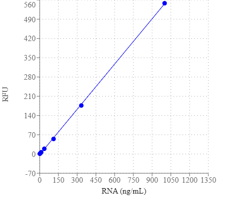
StrandBrite 绿色荧光RNA定量试剂盒是美国AAT Bioquest生产的RNA检测试剂盒,检测和定量少量RNA对于各种分子生物学程序非常重要,例如测量体外转录RNA的产量和测量RNA浓度,然后进行Northern印迹分析,S1核酸酶测定,RNase保护测定,cDNA文库制备,反向转录PCR和差异显示PCR。常用的测量核酸浓度的技术是测定260nm处的吸光度。基于吸光度的方法的主要缺点是由蛋白质,游离核苷酸和其他UV吸收化合物引起的干扰。使用敏感的荧光核酸染色可以缓解这种干扰问题。 StrandBrite RNA定量试剂是一种超灵敏荧光核酸染色剂,用于定量溶液中的RNA。 StrandBrite RNA定量试剂可使用荧光酶标仪或荧光分光光度计检测低至5 ng / mL的RNA。我们的StrandBrite 绿色荧光RNA定量试剂盒包括我们的StrandBrite 绿色核酸染色,具有优化且稳健的方案。它提供了一种方便的方法来量化溶液中的RNA。金畔生物是AAT Bioquest的中国代理商,为您提供优质的StrandBrite 绿色荧光RNA定量试剂盒 。 


适用仪器


荧光酶标仪  
Ex: 490 nm
Em: 545 nm
Cutoff: 515 nm
推荐孔板: 纯黑色孔板
   
实验方案

96孔板检测示例

操作步骤

以下方案是使用StrandBrite Green定量RNA的示例。 打开前让所有组件升温至室温。处理所有材料时,务必使用干净的一次性手套 使用无核酸酶水和无菌一次性聚丙烯塑料制品进行试剂制备。

注意:没有数据可用于解决StrandBrite Green RNA染色的致突变性或毒性。 因为这种试剂与核酸结合,所以应将其作为潜在的诱变剂进行处理,并进行适当的处理。 应谨慎处理DMSO储备溶液,因为已知DMSO有助于有机分子进入组织。


1.准备1X测定缓冲液

通过用无菌,蒸馏,无核酸酶的水稀释浓缩的缓冲液20X(组分B)来制备1X测定缓冲液。


2.准备StrandBrite 绿色工作溶液

通过在1X测定缓冲液中将浓缩的DMSO溶液稀释200倍来制备StrandBrite Green工作溶液。例如,将50μLStrandBrite Green(组分A)加入10 mL 1X测定缓冲液中(来自步骤1)。通过用铝箔覆盖或将其置于黑暗中来保护工作溶液免受光照。

注1:我们建议将此溶液用塑料容器而不是玻璃制备,因为染料可能会吸附到玻璃表面。

注2:为获得佳结果,该溶液应在制备后几小时内使用。


3.准备RNA标准品的连续稀释液(0至1μg/ mL):

3.1将10μL100μg/ mL RNA原液(组分C)加入990μL分析缓冲液(组分B)中,得到1μg/ mL RNA溶液,然后进行1:3连续稀释,得到约1000,300, 100,30,10,3,1和0 ng / mL。

注意:未使用的核糖体RNA标准品(组分C)应在无核酸酶的塑料瓶中分成单次使用的等分试样,并储存在-20℃。

3.2如说明书中表1和2中所述,将RNA标准品和含有测试样品的RNA添加到96孔固体黑色微孔板中。


4.运行RNA测定:

4.1将100μLStrandBrite 绿色工作溶液(来自步骤2)添加到RNA标准品,空白对照和测试样品的每个孔中(参见步骤3)中,使总RNA测定体积为200μL/孔。

注意:对于384孔板,每孔加入25μL样品和25μLStrandBrite 绿色工作溶液。

4.2在室温下孵育反应2至5分钟,避光。

4.3使用荧光分光光度计在Ex / Em = 490 / 545nm(515nm处的截止值)下观察荧光增加。

注意:为尽量减少光漂白,请保持所有样品的荧光测量时间不变。

4.4空白孔中的荧光(仅含测定缓冲液)用作对照,并从具有RNA标准品或测试样品的那些比色皿的值中减去。根据RNA标准曲线确定样品的RNA浓度。


参考文献

Inhibitors of Streptococcus pneumoniae surface endonuclease EndA discovered by high-throughput screening using a PicoGreen fluorescence assay
Authors: Peterson EJ, Kireev D, Moon AF, Midon M, Janzen WP, Pingoud A, Pedersen LC, Singleton SF.
Journal: J Biomol Screen (2013): 247

Validation of a PicoGreen-based DNA quantification integrated in an RNA extraction method for two-dimensional and three-dimensional cell cultures
Authors: Chen Y, Sonnaert M, Roberts SJ, Luyten FP, Schrooten J.
Journal: Tissue Eng Part C Methods (2012): 444

Characterization of PicoGreen interaction with dsDNA and the origin of its fluorescence enhancement upon binding
Authors: Dragan AI, Casas-Finet JR, Bishop ES, Strouse RJ, Schenerman MA, Geddes CD.
Journal: Biophys J (2010): 3010

Comparison of SYBR Green I-, PicoGreen-, and [3H]-hypoxanthine-based assays for in vitro antimalarial screening of plants from Nigerian ethnomedicine
Authors: Abiodun OO, Gbotosho GO, Ajaiyeoba EO, Happi CT, Hofer S, Wittlin S, Sowunmi A, Brun R, Oduola AM.
Journal: Parasitol Res (2010): 933

Metal-enhanced PicoGreen fluorescence: application to fast and ultra-sensitive pg/ml DNA quantitation
Authors: Dragan AI, Bishop ES, Casas-Finet JR, Strouse RJ, Schenerman MA, Geddes CD.
Journal: J Immunol Methods (2010): 95

Quantification of dsDNA using the Hitachi F-7000 Fluorescence Spectrophotometer and PicoGreen dye
Authors: Moreno LA, Cox KL.
Journal: J Vis Exp. (2010)

Development and characterization of a novel host cell DNA assay using ultra-sensitive fluorescent nucleic acid stain “PicoGreen”
Authors: Ikeda Y, Iwakiri S, Yoshimori T.
Journal: J Pharm Biomed Anal (2009): 997

Enhanced DNA dynamics due to cationic reagents, topological states of dsDNA and high mobility group box 1 as probed by PicoGreen
Authors: Noothi SK, Kombrabail M, Kundu TK, Krishnamoorthy G, Rao BJ.
Journal: FEBS J (2009): 541

Factors affecting quantification of total DNA by UV spectroscopy and PicoGreen fluorescence
Authors: Holden MJ, Haynes RJ, Rabb SA, Satija N, Yang K, Blasic JR, Jr.
Journal: J Agric Food Chem (2009): 7221

Label-free DNA sequence detection with enhanced sensitivity and selectivity using cationic conjugated polymers and PicoGreen
Authors: Ren X, Xu QH.
Journal: Langmuir (2009): 43
 

相关产品

产品名称 货号
StrandBrite 绿色荧光RNA定量试剂盒 高选择性 Cat#17657
StrandBrite 绿色荧光RNA定量试剂盒 Cat#17656

StrandBrite 绿色荧光RNA定量试剂盒-AAT Bioquest荧光染料

上海金畔生物科技有限公司代理AAT Bioquest荧光染料全线产品,欢迎访问AAT Bioquest荧光染料官网了解更多信息。
StrandBrite 绿色荧光RNA定量试剂盒价格 4245
产品规格

100 Tests

产品货号

StrandBrite 绿色荧光RNA定量试剂盒

产品参数
Ex (nm) 509 Em (nm) 527
分子量 溶剂
存储条件 在零下15度以下保存, 避免光照
产品概述

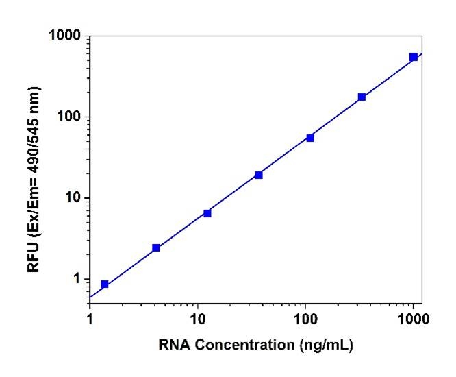
StrandBrite 绿色荧光RNA定量试剂盒是美国AAT Bioquest生产的RNA检测试剂盒,检测和定量少量RNA对于各种分子生物学程序非常重要,例如测量体外转录RNA的产量和测量RNA浓度,然后进行Northern印迹分析,S1核酸酶测定,RNase保护测定,cDNA文库制备,反向转录PCR和差异显示PCR。常用的测量核酸浓度的技术是测定260nm处的吸光度。基于吸光度的方法的主要缺点是由蛋白质,游离核苷酸和其他UV吸收化合物引起的干扰。使用敏感的荧光核酸染色可以缓解这种干扰问题。 StrandBrite RNA定量试剂是一种超灵敏荧光核酸染色剂,用于定量溶液中的RNA。 StrandBrite RNA定量试剂可使用荧光酶标仪或荧光分光光度计检测低至5 ng / mL的RNA。我们的StrandBrite 绿色荧光RNA定量试剂盒包括我们的StrandBrite 绿色核酸染色,具有优化且稳定的方案。它提供了一种方便的方法来量化溶液中的RNA。金畔生物是AAT Bioquest的中国代理商,为您提供优质的StrandBrite 绿色荧光RNA定量试剂盒 。 


适用仪器


分光光度计  
Ex: 490 nm
Em: 545 nm
Cutoff: 515 nm
实验方案

96孔板检测示例

操作步骤

以下方案是使用StrandBrite Green定量RNA的示例。 打开前让所有组件升温至室温。处理所有材料时,务必使用干净的一次性手套 使用无核酸酶水和无菌一次性聚丙烯塑料制品进行试剂制备。

注意:没有数据可用于解决StrandBrite Green RNA染色的致突变性或毒性。 因为这种试剂与核酸结合,所以应将其作为潜在的诱变剂进行处理,并进行适当的处理。 应谨慎处理DMSO储备溶液,因为已知DMSO有助于有机分子进入组织。


1.准备1X测定缓冲液

通过用无菌,蒸馏,无核酸酶的水稀释浓缩的缓冲液20X(组分B)来制备1X测定缓冲液。


2.准备StrandBrite 绿色工作溶液

通过在1X测定缓冲液中将浓缩的DMSO溶液稀释200倍来制备StrandBrite Green工作溶液。例如,将50μLStrandBrite Green(组分A)加入10 mL 1X测定缓冲液中(来自步骤1)。通过用铝箔覆盖或将其置于黑暗中来保护工作溶液免受光照。

注1:我们建议将此溶液用塑料容器而不是玻璃制备,因为染料可能会吸附到玻璃表面。

注2:为获得佳结果,该溶液应在制备后几小时内使用。


3.准备RNA标准品的连续稀释液(0至1μg/ mL):

3.1将10μL100μg/ mL RNA原液(组分C)加入990μL分析缓冲液(组分B)中,得到1μg/ mL RNA溶液,然后进行1:3连续稀释,得到约1000,300, 100,30,10,3,1和0 ng / mL。

注意:未使用的核糖体RNA标准品(组分C)应在无核酸酶的塑料瓶中分成单次使用的等分试样,并储存在-20℃。

3.2如说明书中表1和2中所述,将RNA标准品和含有测试样品的RNA添加到96孔固体黑色微孔板中。


4.运行RNA测定:

4.1将100μLStrandBrite 绿色工作溶液(来自步骤2)添加到RNA标准品,空白对照和测试样品的每个孔中(参见步骤3)中,使总RNA测定体积为200μL/孔。

注意:对于384孔板,每孔加入25μL样品和25μLStrandBrite 绿色工作溶液。

4.2在室温下孵育反应2至5分钟,避光。

4.3使用荧光分光光度计在Ex / Em = 490 / 545nm(515nm处的截止值)下观察荧光增加。

注意:为尽量减少光漂白,请保持所有样品的荧光测量时间不变。

4.4空白孔中的荧光(仅含测定缓冲液)用作对照,并从具有RNA标准品或测试样品的那些比色皿的值中减去。根据RNA标准曲线确定样品的RNA浓度。


参考文献

Inhibitors of Streptococcus pneumoniae surface endonuclease EndA discovered by high-throughput screening using a PicoGreen fluorescence assay
Authors: Peterson EJ, Kireev D, Moon AF, Midon M, Janzen WP, Pingoud A, Pedersen LC, Singleton SF.
Journal: J Biomol Screen (2013): 247

Validation of a PicoGreen-based DNA quantification integrated in an RNA extraction method for two-dimensional and three-dimensional cell cultures
Authors: Chen Y, Sonnaert M, Roberts SJ, Luyten FP, Schrooten J.
Journal: Tissue Eng Part C Methods (2012): 444

Characterization of PicoGreen interaction with dsDNA and the origin of its fluorescence enhancement upon binding
Authors: Dragan AI, Casas-Finet JR, Bishop ES, Strouse RJ, Schenerman MA, Geddes CD.
Journal: Biophys J (2010): 3010

Comparison of SYBR Green I-, PicoGreen-, and [3H]-hypoxanthine-based assays for in vitro antimalarial screening of plants from Nigerian ethnomedicine
Authors: Abiodun OO, Gbotosho GO, Ajaiyeoba EO, Happi CT, Hofer S, Wittlin S, Sowunmi A, Brun R, Oduola AM.
Journal: Parasitol Res (2010): 933

Metal-enhanced PicoGreen fluorescence: application to fast and ultra-sensitive pg/ml DNA quantitation
Authors: Dragan AI, Bishop ES, Casas-Finet JR, Strouse RJ, Schenerman MA, Geddes CD.
Journal: J Immunol Methods (2010): 95

Quantification of dsDNA using the Hitachi F-7000 Fluorescence Spectrophotometer and PicoGreen dye
Authors: Moreno LA, Cox KL.
Journal: J Vis Exp. (2010)

Development and characterization of a novel host cell DNA assay using ultra-sensitive fluorescent nucleic acid stain “PicoGreen”
Authors: Ikeda Y, Iwakiri S, Yoshimori T.
Journal: J Pharm Biomed Anal (2009): 997

Enhanced DNA dynamics due to cationic reagents, topological states of dsDNA and high mobility group box 1 as probed by PicoGreen
Authors: Noothi SK, Kombrabail M, Kundu TK, Krishnamoorthy G, Rao BJ.
Journal: FEBS J (2009): 541

Factors affecting quantification of total DNA by UV spectroscopy and PicoGreen fluorescence
Authors: Holden MJ, Haynes RJ, Rabb SA, Satija N, Yang K, Blasic JR, Jr.
Journal: J Agric Food Chem (2009): 7221

Label-free DNA sequence detection with enhanced sensitivity and selectivity using cationic conjugated polymers and PicoGreen
Authors: Ren X, Xu QH.
Journal: Langmuir (2009): 43
 

相关产品

产品名称 货号
StrandBrite 绿色荧光RNA定量试剂盒 高选择性 Cat#17657
StrandBrite 绿色荧光RNA定量试剂盒 Cat#17656

StrandBrite 绿色荧光RNA定量试剂盒 高选择性-AAT Bioquest荧光染料

上海金畔生物科技有限公司代理AAT Bioquest荧光染料全线产品,欢迎访问AAT Bioquest荧光染料官网了解更多信息。
StrandBrite 绿色荧光RNA定量试剂盒 高选择性价格 4957
产品规格

100 Tests

产品货号

StrandBrite 绿色荧光RNA定量试剂盒 高选择性

产品参数
Ex (nm) 509 Em (nm) 527
分子量 溶剂
存储条件
产品概述

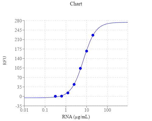
StrandBrite 绿色荧光RNA定量试剂盒是美国AAT Bioquest生产的RNA检测试剂盒,分析活细胞中 RNA 的主要挑战是 DNA 引起的干扰。为了解决这些困难,AAT Bioquest 开发了 StrandBrite™ RNA Green,这是一种出色的 RNA 选择性探针,在与 RNA 结合后可产生显着增强的绿色荧光。它已成功用于活细胞的流式细胞分析。 StrandBrite™ RNA Green 很容易进入活细胞。它的激发/发射波长为490/540 nm。在DNase消化测试中,用StrandBrite RNA Green染色的固定细胞中没有观察到荧光强度的显着变化。相反,RNase 消化后,初始荧光信号立即减弱。这些结果表明,初始荧光信号是由 StrandBrite RNA Green 与细胞中 RNA 的特异性相互作用产生的。活细胞短暂暴露于抗霉素 D 确实会在药物去除后 6 小时内以剂量依赖性方式抑制 RNA 合成。这些数据表明 StrandBrite RNA Green 是一种灵敏的 RNA 选择性染料,用于对活细胞和固定细胞中的核仁 RNA 进行染色。 StrandBrite RNA Green 比常用的 SYTO® RNASelect™ 染料具有更少的 DNA 干扰。 StrandBrite™ RNA Green 是一种高度 RNA 选择性荧光探针。由于其优异的细胞通透性和光谱特性,已成功用于活细胞中的流式细胞术RNA分析和荧光显微镜。它可以用 488 nm 蓝色激光很好地激发并在 FITC 通道中进行监测。 StrandBrite™ RNA Green 提供了一种通过单个孵育步骤识别和标记细胞的有价值的方法,并且可以比常用的 SYTO® RNASelect™ 具有更好的选择性区分 RNA 和 DNA。金畔生物是AAT Bioquest的中国代理商,为您提供优质的StrandBrite 绿色荧光RNA定量试剂盒 。 


适用仪器


荧光酶标仪  
Ex: 490 nm
Em: 540 nm
Cutoff: 515 nm
推荐孔板: 纯黑色孔板
   
实验方案

96孔板检测示例

操作步骤

以下方案是使用StrandBrite Green定量RNA的示例。 打开前让所有组件升温至室温。处理所有材料时,务必使用干净的一次性手套 使用无核酸酶水和无菌一次性聚丙烯塑料制品进行试剂制备。

注意:没有数据可用于解决StrandBrite Green RNA染色的致突变性或毒性。 因为这种试剂与核酸结合,所以应将其作为潜在的诱变剂进行处理,并进行适当的处理。 应谨慎处理DMSO储备溶液,因为已知DMSO有助于有机分子进入组织。


1.准备1X测定缓冲液

通过用无菌,蒸馏,无核酸酶的水稀释浓缩的缓冲液20X(组分B)来制备1X测定缓冲液。


2.准备StrandBrite 绿色工作溶液

通过在1X测定缓冲液中将浓缩的DMSO溶液稀释200倍来制备StrandBrite Green工作溶液。例如,将50μLStrandBrite Green(组分A)加入10 mL 1X测定缓冲液中(来自步骤1)。通过用铝箔覆盖或将其置于黑暗中来保护工作溶液免受光照。

注1:我们建议将此溶液用塑料容器而不是玻璃制备,因为染料可能会吸附到玻璃表面。

注2:为获得佳结果,该溶液应在制备后几小时内使用。


3.准备RNA标准品的连续稀释液(0至1μg/ mL):

3.1将10μL100μg/ mL RNA原液(组分C)加入990μL分析缓冲液(组分B)中,得到1μg/ mL RNA溶液,然后进行1:3连续稀释,得到约1000,300, 100,30,10,3,1和0 ng / mL。

注意:未使用的核糖体RNA标准品(组分C)应在无核酸酶的塑料瓶中分成单次使用的等分试样,并储存在-20℃。

3.2如说明书中表1和2中所述,将RNA标准品和含有测试样品的RNA添加到96孔固体黑色微孔板中。


4.运行RNA测定:

4.1将100μLStrandBrite 绿色工作溶液(来自步骤2)添加到RNA标准品,空白对照和测试样品的每个孔中(参见步骤3)中,使总RNA测定体积为200μL/孔。

注意:对于384孔板,每孔加入25μL样品和25μLStrandBrite 绿色工作溶液。

4.2在室温下孵育反应2至5分钟,避光。

4.3使用荧光分光光度计在Ex / Em = 490 / 545nm(515nm处的截止值)下观察荧光增加。

注意:为尽量减少光漂白,请保持所有样品的荧光测量时间不变。

4.4空白孔中的荧光(仅含测定缓冲液)用作对照,并从具有RNA标准品或测试样品的那些比色皿的值中减去。根据RNA标准曲线确定样品的RNA浓度。


参考文献

Inhibitors of Streptococcus pneumoniae surface endonuclease EndA discovered by high-throughput screening using a PicoGreen fluorescence assay
Authors: Peterson EJ, Kireev D, Moon AF, Midon M, Janzen WP, Pingoud A, Pedersen LC, Singleton SF.
Journal: J Biomol Screen (2013): 247

Validation of a PicoGreen-based DNA quantification integrated in an RNA extraction method for two-dimensional and three-dimensional cell cultures
Authors: Chen Y, Sonnaert M, Roberts SJ, Luyten FP, Schrooten J.
Journal: Tissue Eng Part C Methods (2012): 444

Characterization of PicoGreen interaction with dsDNA and the origin of its fluorescence enhancement upon binding
Authors: Dragan AI, Casas-Finet JR, Bishop ES, Strouse RJ, Schenerman MA, Geddes CD.
Journal: Biophys J (2010): 3010

Comparison of SYBR Green I-, PicoGreen-, and [3H]-hypoxanthine-based assays for in vitro antimalarial screening of plants from Nigerian ethnomedicine
Authors: Abiodun OO, Gbotosho GO, Ajaiyeoba EO, Happi CT, Hofer S, Wittlin S, Sowunmi A, Brun R, Oduola AM.
Journal: Parasitol Res (2010): 933

Metal-enhanced PicoGreen fluorescence: application to fast and ultra-sensitive pg/ml DNA quantitation
Authors: Dragan AI, Bishop ES, Casas-Finet JR, Strouse RJ, Schenerman MA, Geddes CD.
Journal: J Immunol Methods (2010): 95

Quantification of dsDNA using the Hitachi F-7000 Fluorescence Spectrophotometer and PicoGreen dye
Authors: Moreno LA, Cox KL.
Journal: J Vis Exp. (2010)

Development and characterization of a novel host cell DNA assay using ultra-sensitive fluorescent nucleic acid stain “PicoGreen”
Authors: Ikeda Y, Iwakiri S, Yoshimori T.
Journal: J Pharm Biomed Anal (2009): 997

Enhanced DNA dynamics due to cationic reagents, topological states of dsDNA and high mobility group box 1 as probed by PicoGreen
Authors: Noothi SK, Kombrabail M, Kundu TK, Krishnamoorthy G, Rao BJ.
Journal: FEBS J (2009): 541

Factors affecting quantification of total DNA by UV spectroscopy and PicoGreen fluorescence
Authors: Holden MJ, Haynes RJ, Rabb SA, Satija N, Yang K, Blasic JR, Jr.
Journal: J Agric Food Chem (2009): 7221

Label-free DNA sequence detection with enhanced sensitivity and selectivity using cationic conjugated polymers and PicoGreen
Authors: Ren X, Xu QH.
Journal: Langmuir (2009): 43
 

相关产品

产品名称 货号
StrandBrite 绿色荧光RNA定量试剂盒 高选择性 Cat#17657
StrandBrite 绿色荧光RNA定量试剂盒 Cat#17656